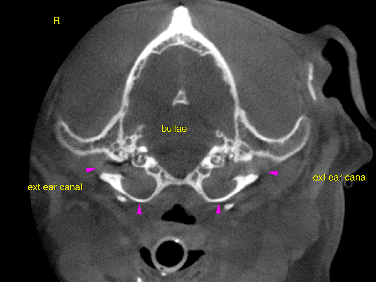
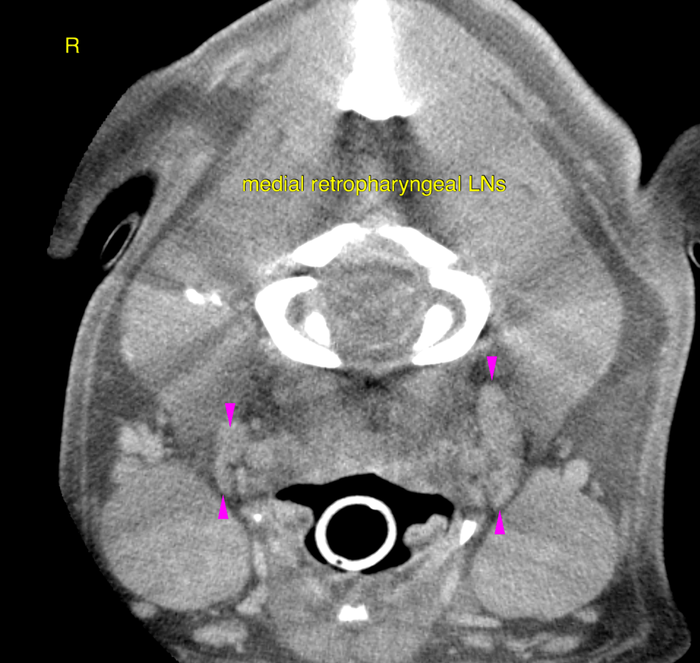
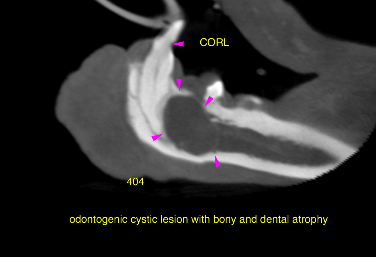
CT of the head, plain and post contrast – The tympanic bullae are completely filled with hypoattenuating material which does not show any contrast enhancement. The material extends into the medial aspect of the ear canal respectively. The wall of the ear canals presents mild generalized thickening with focal small bony metaplasia. The general conformation of the horizontal part of the ear canals is narrow and tapering towards the entrance into the bulla. Mild generalized thickening of both bulla walls is noted. There is an expansile cystic lesion of 2 cm diameter emerging from the tooth root 404. The lesion is space occupying and causes bone & dental atrophy as well as a soft tissue swelling. Multifocal odontoclastic (CORL) lesions are seen at the tooth necks and crowns. The left medial retropharyngeal lymph nodes presents moderate symmetrical enlargement with mildly heterogenous contrast enhancement.