CT – Abscess with cellulitis and reactive lymphadenitis in a 3 year old M Irish Setter dog

Case Study

CT – Abscess with cellulitis and reactive lymphadenitis in a 3 year old M Irish Setter dog

The patient is a 3 year old M Irish Setter dog presented for a mass on the nec of 3-4 weeks duration. Cytology indicated evidence of abscess but the mass has continued to enlarge despite antibiotic therapy and NSAIDS.

Physical exam: grapefruit-sized mass on neck

The patient is a 3 year old M Irish Setter dog presented for a mass on the nec of 3-4 weeks duration. Cytology indicated evidence of abscess but the mass has continued to enlarge despite antibiotic therapy and NSAIDS.

Physical exam: grapefruit-sized mass on neck

Image Interpretation

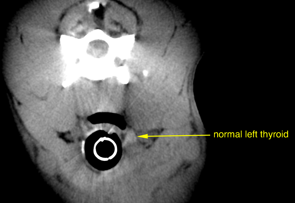
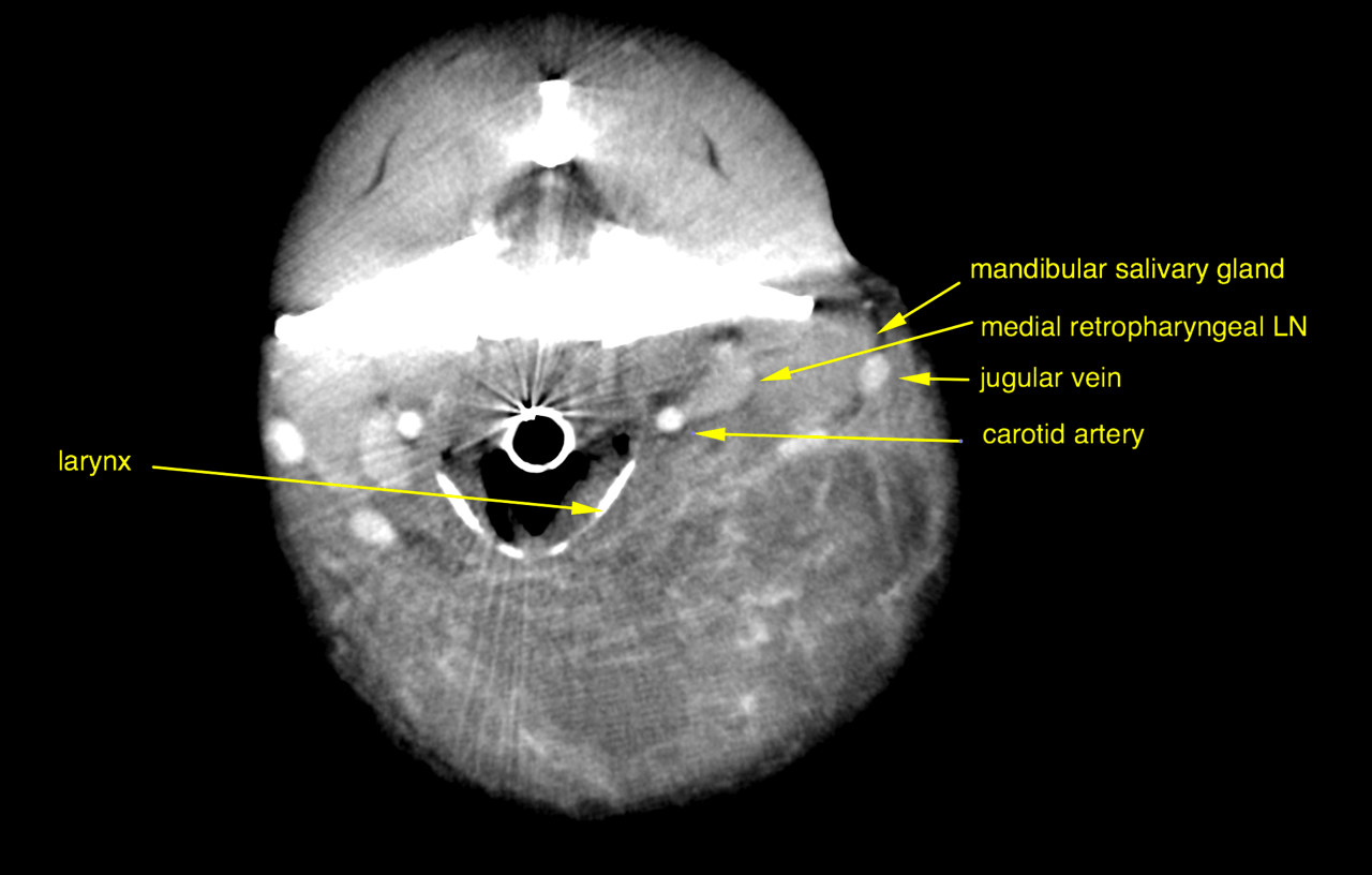
CT of the neck, plain and post contrast – The computed tomography reveals a soft tissue attenuating ovoid mass lesion in the
left ventral neck next to the trachea, larynx, hyoid bone and base of the tongue. The
mass is located ventral and lateral to the trachea and larynx and ventral to the
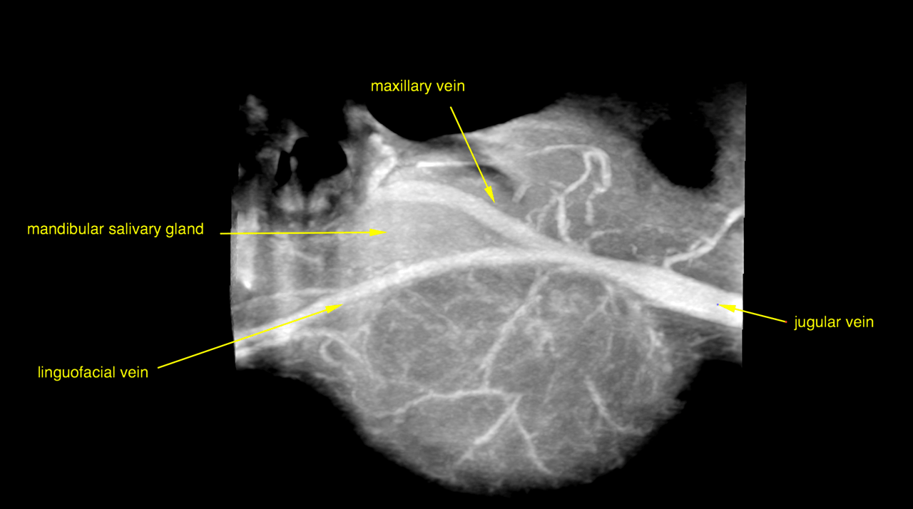
mandibular salivary gland and medial retropharyngeal lymph node. Furthermore it is
ventral to the jugular, maxillary and linguofacial veins as well as ventral to the
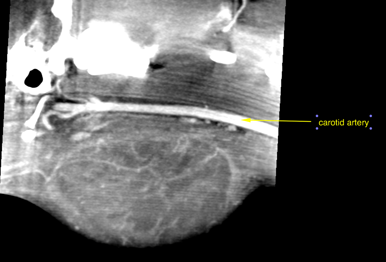
common carotid artery and carotid bifurcation.
The mass is in close proximity to the left mandibular salivary gland and sublingual
salivary gland but a connection with these is not explicitly seen.
Foreign material is not seen within the mass.
The size of the lesion is 8 x 8 x 7 cm. It exerts a marked mass effect on the surrounding
anatomy with rightward tracheal and laryngeal displacement.
The mass has a layered appearance with fat stranding in the periphery. The capsule of
the mass reveals thick and irregular rim enhancement, multifocal septae in the outer
layers of the mass enhance contrast whereas the center and ventral aspect of the mass
spares contrast. Multifocal small feeding vessels are seen in the periphery of the mass.
The ipsilateral medial retropharyngeal lymph node and both mandibular lymph centers
show signs of reactive hyperplasia.

DX

Abscess with surrounding cellulitis and reactive lymphadenitis

Outcome

Surgical therapy is advised
Foreign material was not seen. Note that this does not rule out hydrolyzed/macerated
organic foreign material entirely as this acquires properties of the surrounding tissue
over time.
Critical neighboring structures include the larynx, laryngeal and vagal nerves, jugular
vein with tributaries and carotid artery with branchings.
An inflamed mucocele or necrotizing tumor are considered unlikely. Yet examination
of the mass content on presence of saliva and histopathological examination of a tissue
sample should be enforced at the time of surgery.

Patient Information

Patient Name : Flynn Michels
Gender : Male, Intact
Species : Canine
Type of Imaging : Ultrasound
Status : Complete
Liz Wuz Here : Yes

Exam Finding

  • Masses

Images

bildschirmfoto_2015-12-18_um_09bildschirmfoto_2015-12-18_um_10bildschirmfoto_2015-12-18_um_10bildschirmfoto_2015-12-18_um_10
Skip to content