A 9-year-old M English Bulldog was presented for weakness. Murmur was difficult to auscultate due to breed conformation.
A 9-year-old M English Bulldog was presented for weakness. Murmur was difficult to auscultate due to breed conformation.
A 9-year-old M English Bulldog was presented for weakness. Murmur was difficult to auscultate due to breed conformation.
A 9-year-old M English Bulldog was presented for weakness. Murmur was difficult to auscultate due to breed conformation.
Mitral insufficiency, myocardial insufficiency, tricuspid insufficiency and pulmonary hypertension. Left and right sided congestive heart failure.
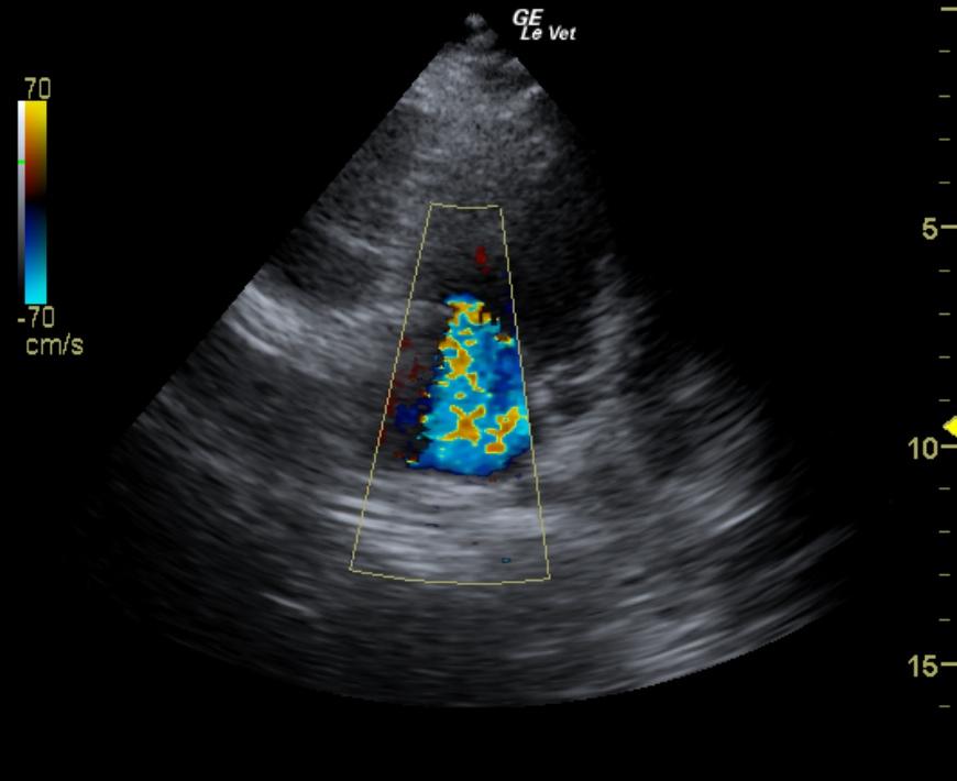
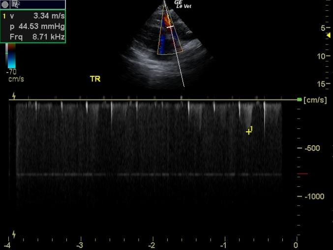
The echocardiogram for this dog presented excessive left atrial size expressed both in the LA/AO and LA max measurements. Left atrial content was anechoic and no evidence of “smoke” or thrombotic activity was noted. The atrial septum was deviated due to volume overload. The mitral valve apparatus demonstrated insufficiency on color flow and spectral Doppler. End point to septal separation was excessive owing to subnormal left ventricular contractility. The left ventricle demonstrated excessive volume (LVIDd measurement below). Ventricular function was subnormal expressed by the fractional shortening. The tricuspid valve presented moderate insufficiency. This is consistent with pulmonary hypertension with prominent right atrial enlargement. The right ventricle demonstrated subnormal kinesis. Rapid assessment of the hepatic veins and vena cava revealed no evidence of passive congestion. This presentation is most consistent AV valvular insufficiency and myocardial insufficiency (subnormal contractility in light of valvular insufficiency) with left sided volume overload and early congestive heart failure. Systemic factors such as hypothyroidism or systemic disease which negatively influence the myocardial contractility may also be playing a role in the presentation. Tricuspid insufficiency velocity was measured at 3.34 m/sec.
The patient was treated with Lasix, Ace inhibitor, Pimobendan, and aspirin. A recheck ultrasound within 2-4 weeks with follow-up renal values and gradual introduction to a geriatric diet was also advised.
Given that hepatic veins were not dilated, the right heart was compensated without evidence of right sided failure even though pulmonary hypertension was present.
Episodes of weakness: cardiovascular disease, respiratory disease, metabolic disease.
None.