A 10-year-old SF Rottweiler cross was presented with a history of lethargy and not doing right. On physical examination depression and polypnea was evident. Abnormalities on serum biochemistry were elevated liver enzyme activity. Cardiomegaly was present on survey radiographs. Blood pressure was normal (140).
A 10-year-old SF Rottweiler cross was presented with a history of lethargy and not doing right. On physical examination depression and polypnea was evident. Abnormalities on serum biochemistry were elevated liver enzyme activity. Cardiomegaly was present on survey radiographs. Blood pressure was normal (140).
Case Study
Cardiac Mass in Right Auricle with Tamponade in a 10 year old Rottweiler Dog
Sonographic Differential Diagnosis
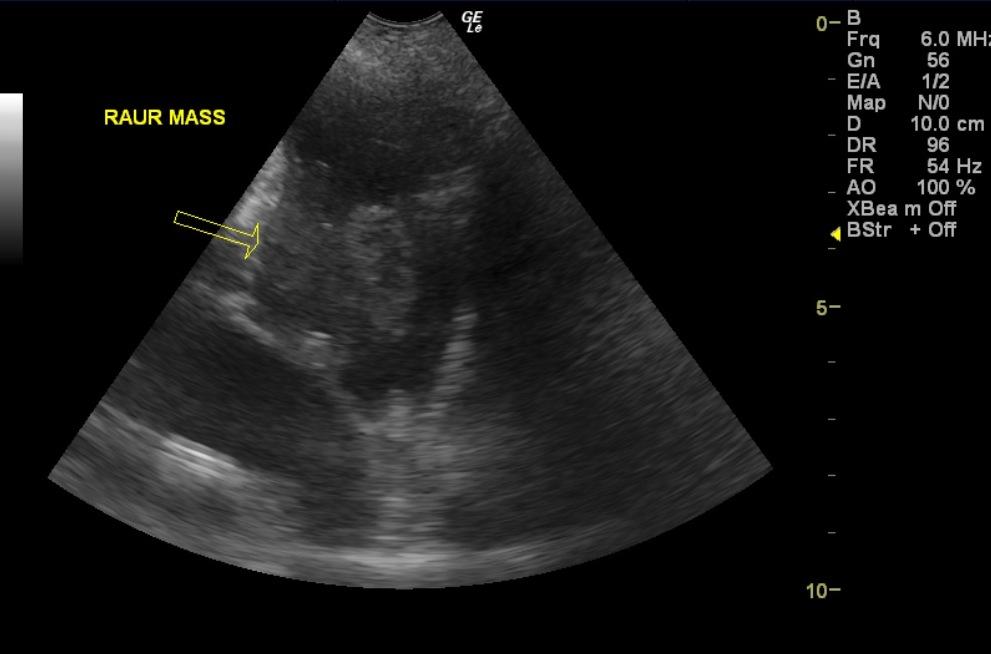
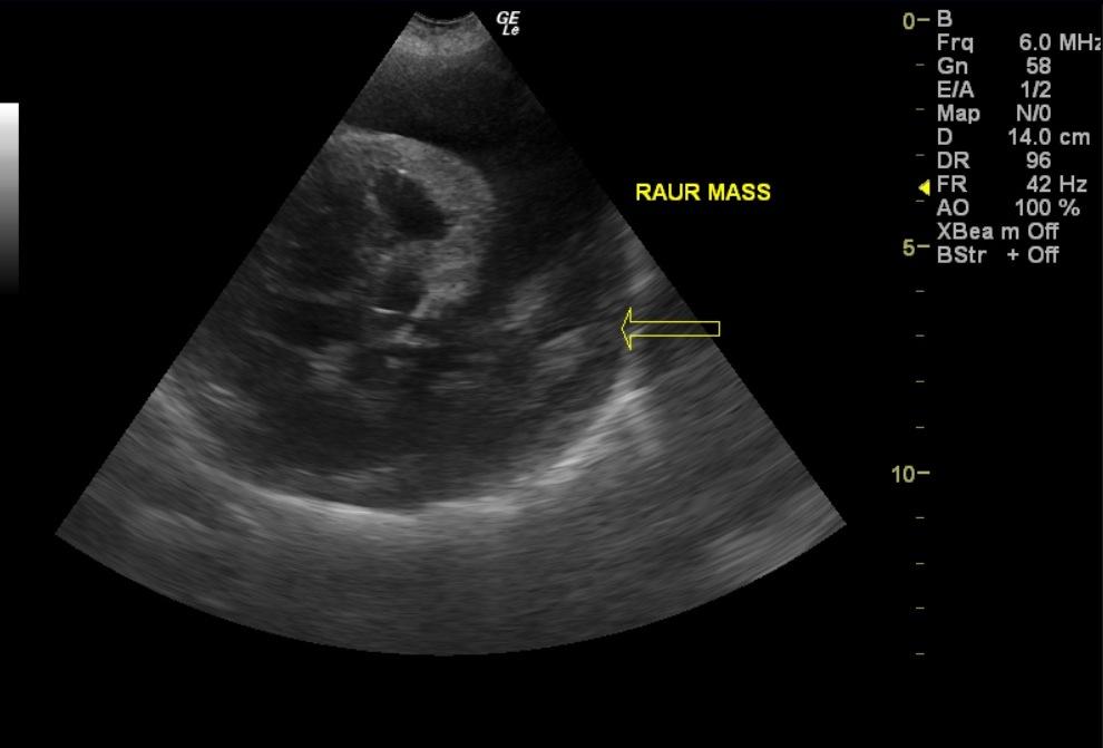
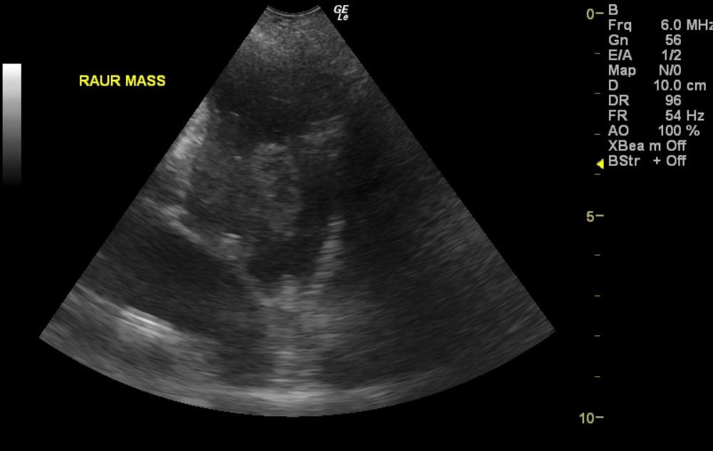
Cardiac mass, right auricle.
Mild to moderate pericardial effusion occupying the contour of the right heart. Further pericardial drainage could be performed in this patient. Given the position of the mass and the echogenicity along with the breed predisposition this is likely hemangiosarcoma. Empirical Adriamycin or empirical protocol for hemangiosarcoma could be considered as an off label rescue attempt with sonographic reassessment after 1 week of therapy. Guarded prognosis.
Image Interpretation
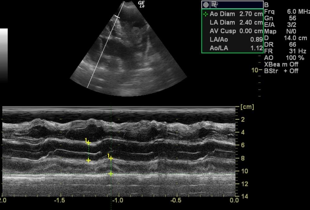
The cardiac presentation in this patient presented mildly subnormal left atrial and left ventricular volume. Contractility was adequate. The left ventricular outflow tract was unremarkable. A 4.5 cm, irregular, hypoechoic mass was present deriving from the right ventricular-right atrial free wall. Right auricular mass occupied pericardial space. There was mild to moderate amount of pericardial effusion noted. This largely occupied the right side of the heart with minimal pericardial effusion along the left side of the heart. This is most consistent with hemangiosarcoma or similar neoplasia given the position and hypoechogenicity.
Clinical Differential Diagnosis
Cardiac -cardiomyopathy (dilated/hypertrophic), pericardial effusion Liver – vacuolar hepatopathy, nodular regeneration, neoplasia, toxins, infectious (viral/bacterial/fungal)
Sampling
None
Video
Patient Information
Patient Name :
Lady D
Gender :
Female, Spayed
Species :
Canine
Type of Imaging : Ultrasound
Status :
Complete
Liz Wuz Here :
Yes
Code :
15-00134
Clinical Signs
- "Not Doing Right"
- Anemia
- Ataxia
- Collapse
- Decreased mobility
- Difficulty walking
- Exercise intolerance
- Lethargy
- Panting
- Weakness
Exam Finding
- Depression
- Lethargy
- Muffled Heart Sounds
- Pale Mucous Membranes
- Panting
- Respiratory Distress
- Tachypnea
- Thready pulses
- Weakness
Images
Blood Chemistry
- Elevated Liver Enzymes
CBC
- Hematocrit, Low
Clinical Signs
- "Not Doing Right"
- Anemia
- Ataxia
- Collapse
- Decreased mobility
- Difficulty walking
- Exercise intolerance
- Lethargy
- Panting
- Weakness