CT of the head –
The computed tomography reveals 2 mass lesions.
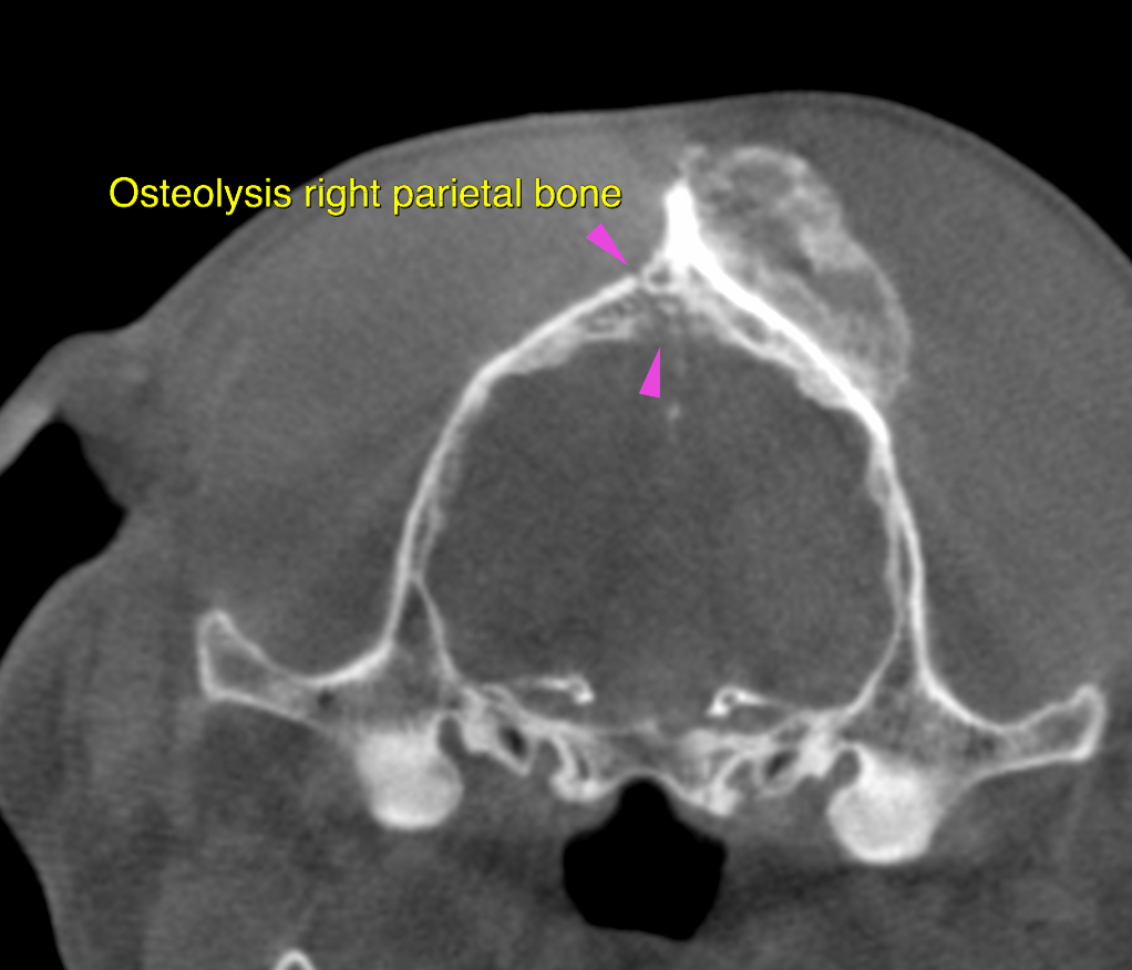
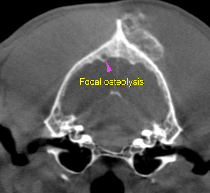
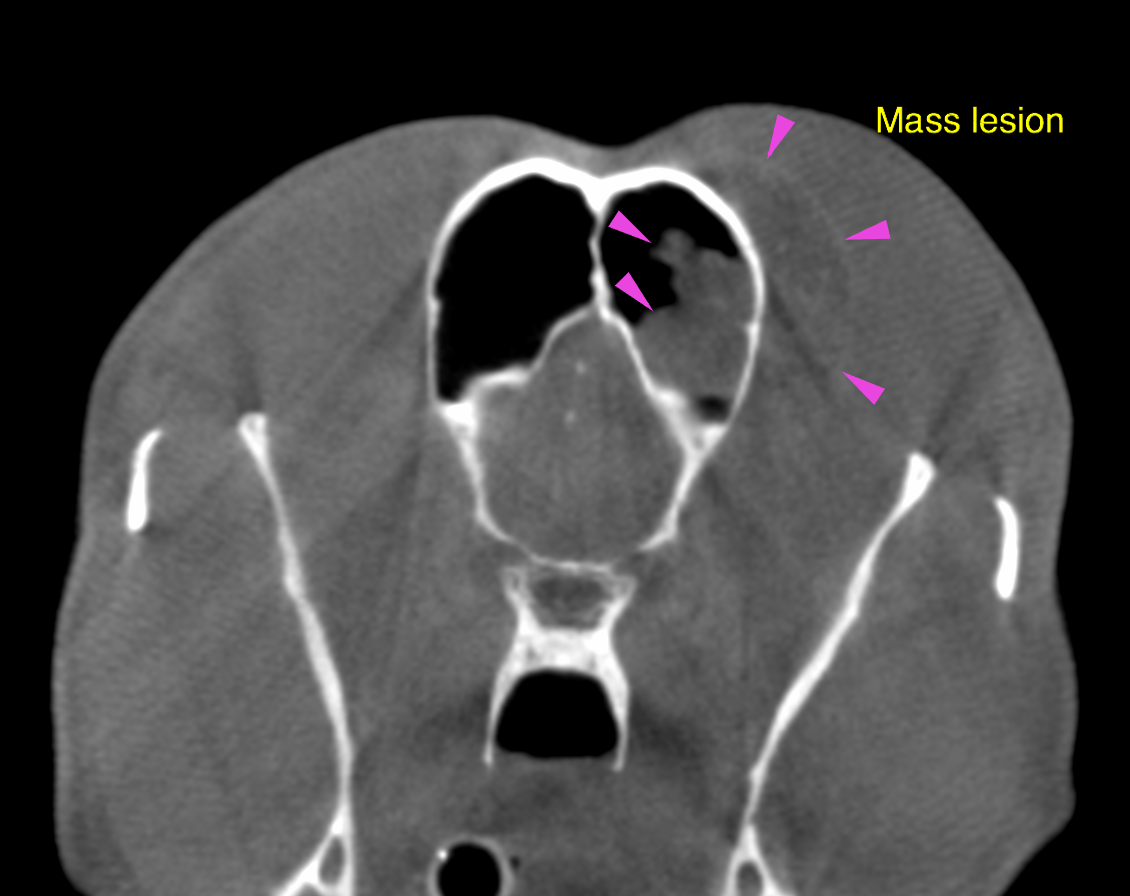
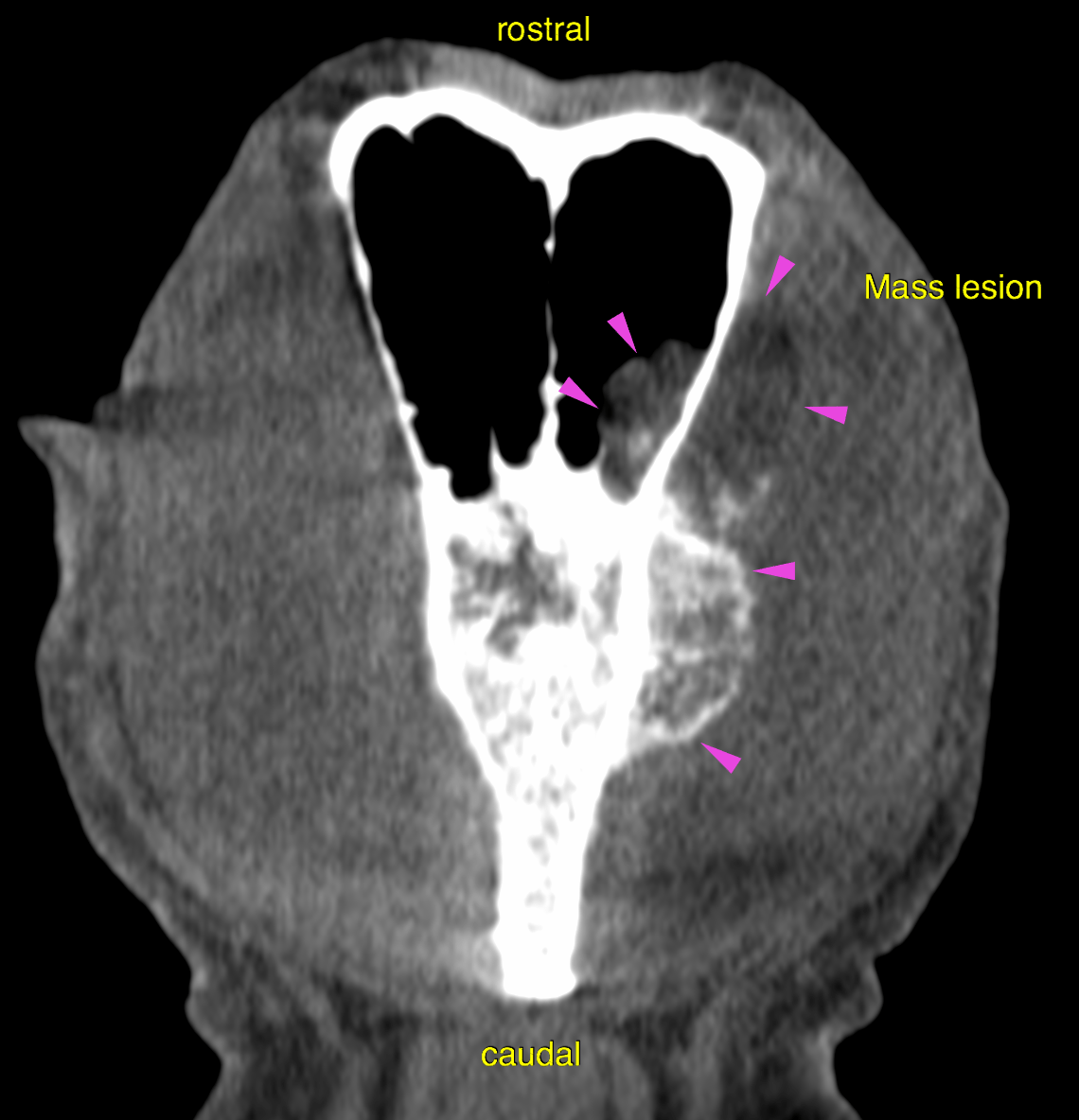
The first mass lesion is emerging from the calvarial bone to the left side of the external occipital protuberance with rostral extension to the frontal bione and sinus. The mass measures approximately 8x3x2cm including the soft tissue component within the masticatory muscles superficial to the mass. Multifocal amorphous periosteal mineralization is seen, at the level of the frontal sinus the mass lesion is non-mineralized and mildly hypoattenuating to the surrounding musculature. The right parietal bone shows focal permeative osteolysis close to the midline level with the parietal lobe of the brain. Complete perforation of the calvarium is not evident yet.
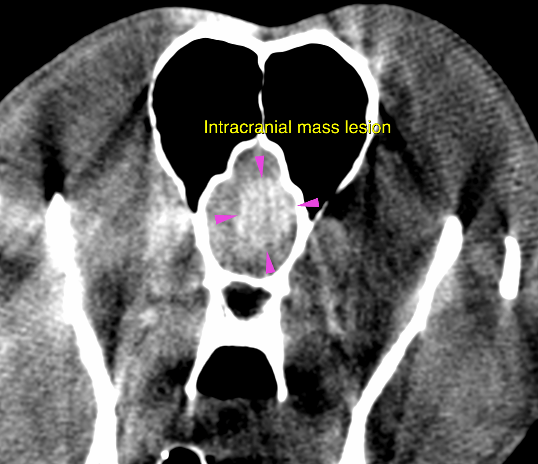
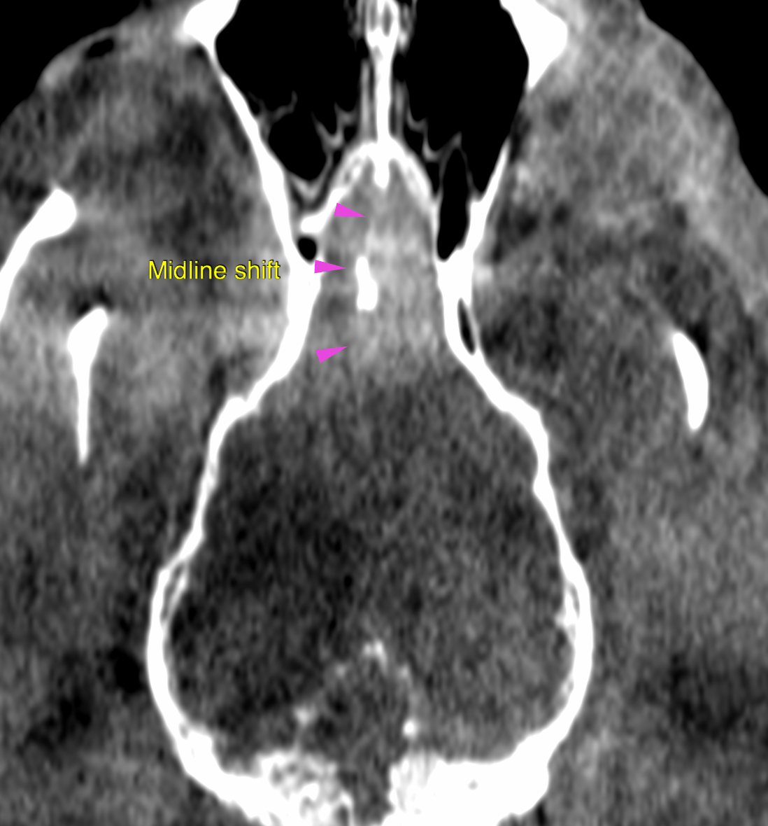
After contrast administration a second mass lesion is seen in an intracranial and extraaxial position emerging from the cerebral falx in the region of the left olfactory bulb. The mass lesion is broadly attached to the frontal bone and leads to a midline shift of the medial rhinal sulcus to the right side. It measures 2x1x1.5cm. There are mineral attenuating small foci at the medial aspect of the cerebral falx.