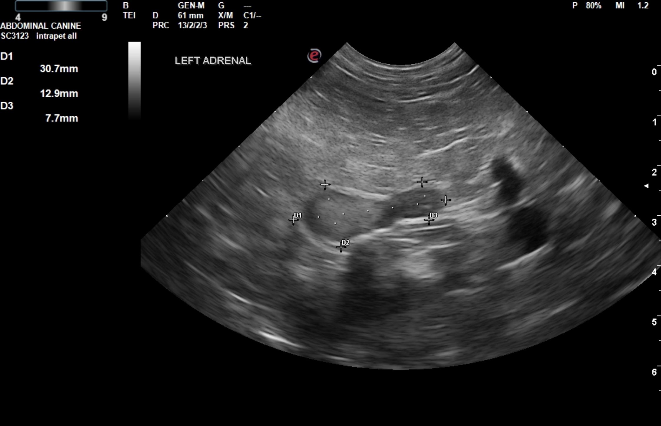
A 15-year-old MN Australian Cattle dog was presented for evaluation of hematuria and weight loss. Urinalysis showed SG of 1.012, proteinuria, and hematuria. The only abnormality on serum biochemistry was mildly elevated ALP (174) activity. On survey radiographs, a soft tissue calcified opacity within the urinary bladder was evident.
A 15-year-old MN Australian Cattle dog was presented for evaluation of hematuria and weight loss. Urinalysis showed SG of 1.012, proteinuria, and hematuria. The only abnormality on serum biochemistry was mildly elevated ALP (174) activity. On survey radiographs, a soft tissue calcified opacity within the urinary bladder was evident.