A 9-year-old intact female Brittany Spaniel dog, with previous history of cystotomy, was presented on emergency at a 24-hour facility for vomiting, shaking, painful abdomen, and extreme lethargy. Physical examination found the patient whining and crying, depressed, with tacky mucous membranes, and a firm distended abdomen. The patient was ambulatory but was most comfortable in sternal recumbency. Abnormalities on CBC and blood chemistry included leukocytosis, increased BUN, and mild hypoalbuminemia. Coagulation panel was within normal range.
A 9-year-old intact female Brittany Spaniel dog, with previous history of cystotomy, was presented on emergency at a 24-hour facility for vomiting, shaking, painful abdomen, and extreme lethargy. Physical examination found the patient whining and crying, depressed, with tacky mucous membranes, and a firm distended abdomen. The patient was ambulatory but was most comfortable in sternal recumbency. Abnormalities on CBC and blood chemistry included leukocytosis, increased BUN, and mild hypoalbuminemia. Coagulation panel was within normal range. Survey radiographs showed generalized loss of detail (consistent with free fluid) and a linear radiodensity in the cranial abdomen. Despite being a poor surgical candidate, the patient was recommended for immediate exploratory surgery due to concern of a ruptured pyometra.
Case Study
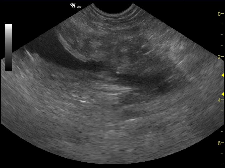
Bladder mass in a 9 year old FI Brittany Spaniel
Sonographic Differential Diagnosis
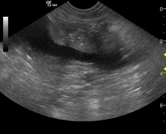
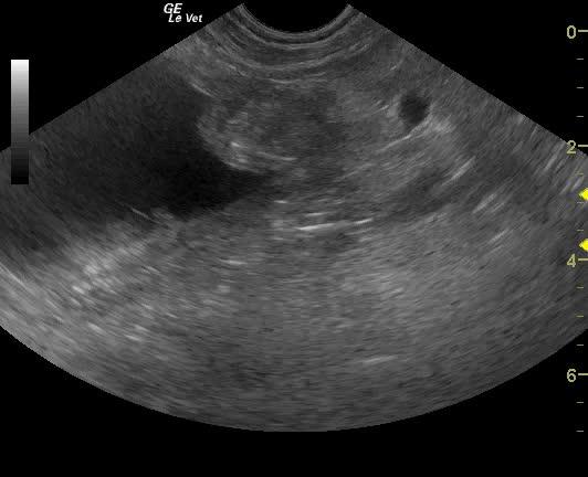
Trigonal bladder mass causing obstruction of the left ureter. Mild to moderate interstitial nephrosis pattern.
Image Interpretation
The kidneys presented a chronic interstitial nephrosis pattern with hyperechoic cortices. Minor hydronephrosis of the left kidney was noted, and was secondary to the obstruction of the ureter at the level of the trigone by an annular mass. This mass appeared to involve the dorsal trigonal area, wrap around the lateral aspect of the bladder, and drain to the cystourethral junction. The mass itself was mineralizing and measured 4.6 x 2.5 cm. The apical, dorsal aspect of the bladder presented a minor noninflamed polypoid change that is consistent with previous inversion from cystotomy. No evidence of rupture was noted.
DX
Outcome
The patient was referred for ultrasound-guided laser ablation of the bladder mass in an effort to liberate the ureter. A stent placement was also discussed as possibility. Following normal thoracic radiographs, an exploratory surgery was performed. A 2-3cm spherical, irregular red bleeding mass was seen adhered to the mucosa within a ruptured bladder. The mass was excised down to the bladder mucosa and the area of bladder rupture was debrided and closed. A Foley catheter was placed. The patient was treated with Piroxicam and Buprenex. The patient was presented a few days later for lethargy, diarrhea, vomiting, and not urinating well. Physical examination found the patient febrile at 102.9 degrees, tachypneic, and depressed. The left aspect of the caudal incision was swollen, erythematous, and inflamed. A yellowish-tan, bloody discharge was draining from the caudal aspect of the incision. No significant abnormalities were seen on CBC or blood chemistry. PCV/TP was 36/8.0. Culture from the incision yielded growth of MRSA. The patient was treated with I.V. fluids, Ampicillin, Baytril, Cerenia, Carafate, Buprenex, Famotidine, and Tramadol. The patient underwent another exploratory surgery in which a draining tract was found following up to the right 4th mammary gland. The patient recovered without event and was discharged with Antirobe.
Clinical Differential Diagnosis
Ascites – modified transudate (neoplasia, splenic torsion), septic exudate (bacterial peritonitis), blood (spleen/liver), urine (bladder/ureter), bile (gall bladder/bile duct).
Sampling
None
Video
Patient Information
Clinical Signs
- Abdominal Pain
- Lethargy
- Shaking
- Vomiting
Exam Finding
- Abdominal Distension
- Dehydration
- Depression
- Vocalizing
Images

Blood Chemistry
- Albumin, Low
- BUN high
CBC
- WBC, High
Clinical Signs
- Abdominal Pain
- Lethargy
- Shaking
- Vomiting