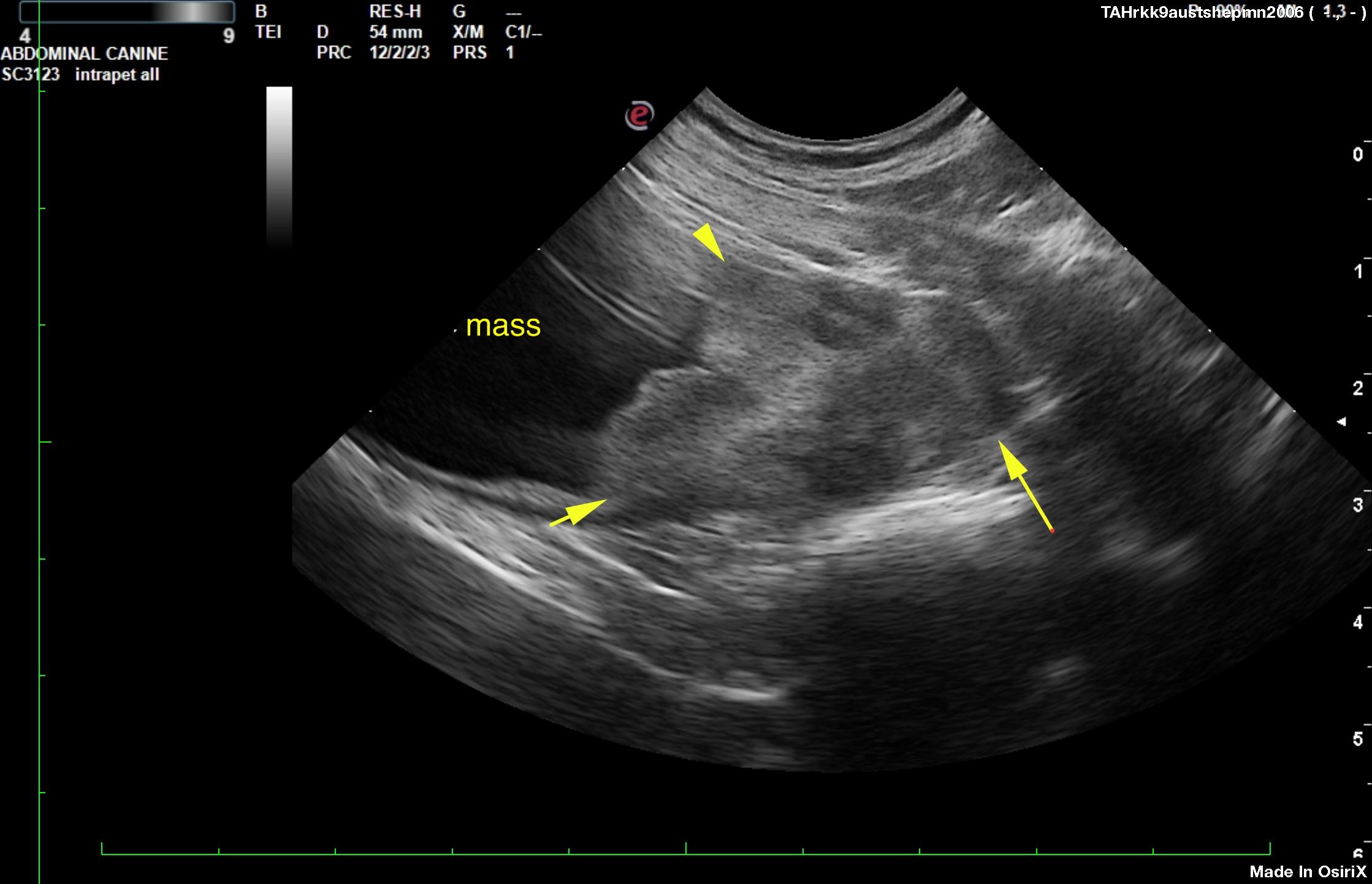
An 11-year-old MN Australian shepherd was presented for evaluation of progressive polyuria and stranguria. Urinalysis showed abnormal epithelial cells. On survey radiographs, mineralization near the pelvis in area of bladder was evident.
An 11-year-old MN Australian shepherd was presented for evaluation of progressive polyuria and stranguria. Urinalysis showed abnormal epithelial cells. On survey radiographs, mineralization near the pelvis in area of bladder was evident.