Bilateral thyroid masses with probable liver metastasis in a 14 year old FS Pug

Case Study

Bilateral thyroid masses with probable liver metastasis in a 14 year old FS Pug

A 14-year-old female spayed Pug with a history of thyroid carcinoma was presented for evaluation of a large mass that extended from her right ear to the thoracic inlet. 
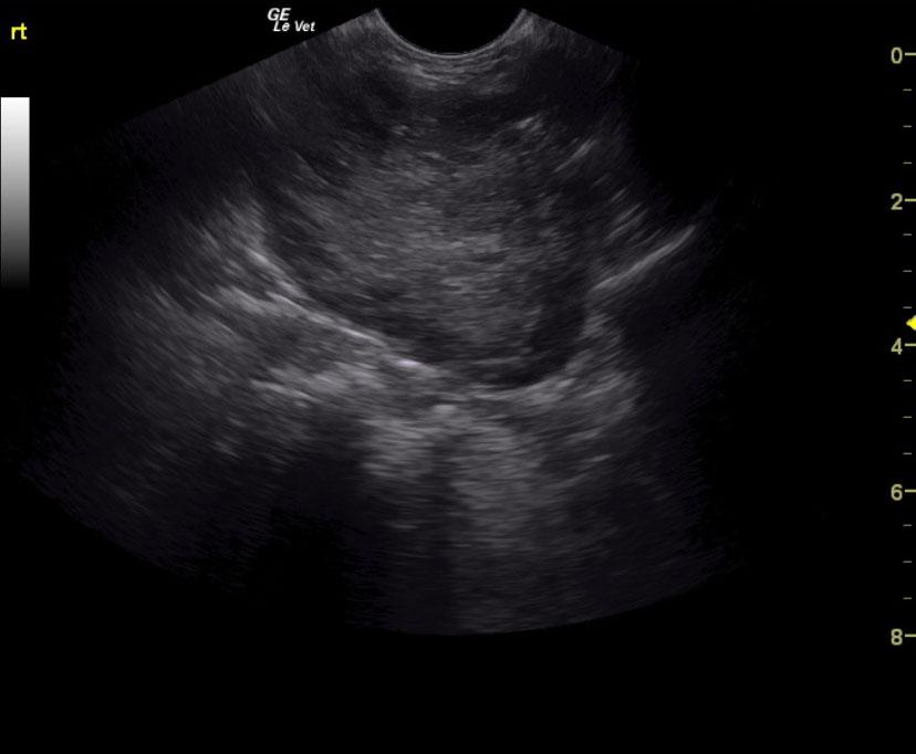
A 14-year-old female spayed Pug with a history of thyroid carcinoma was presented for evaluation of a large mass that extended from her right ear to the thoracic inlet. 
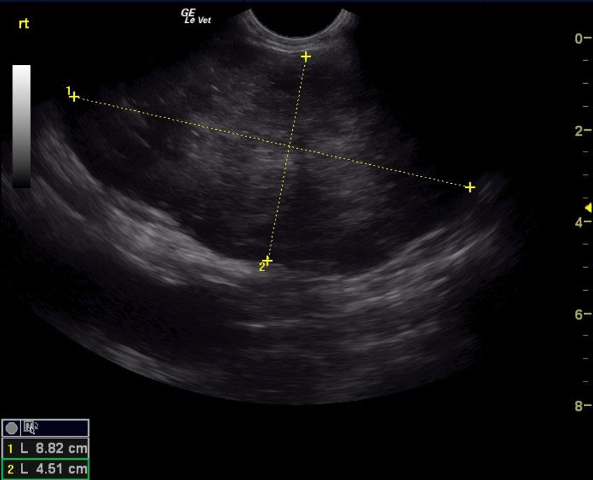
Sonographic Differential Diagnosis

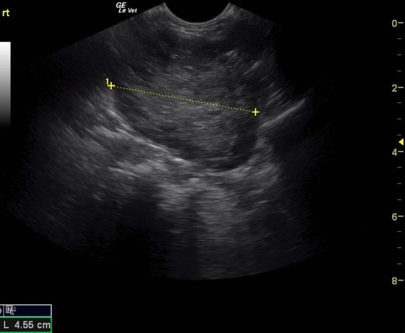
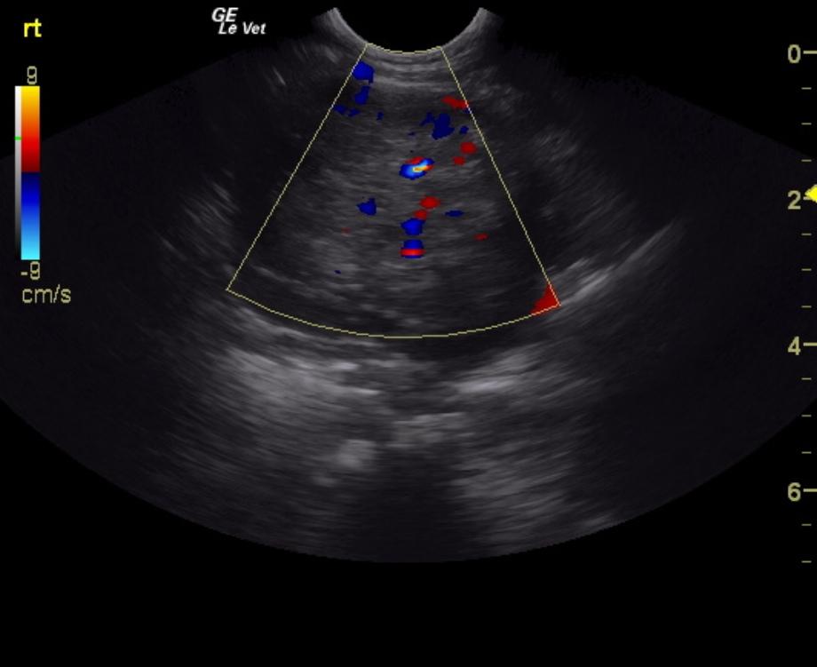
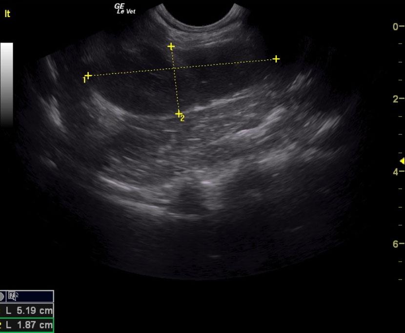
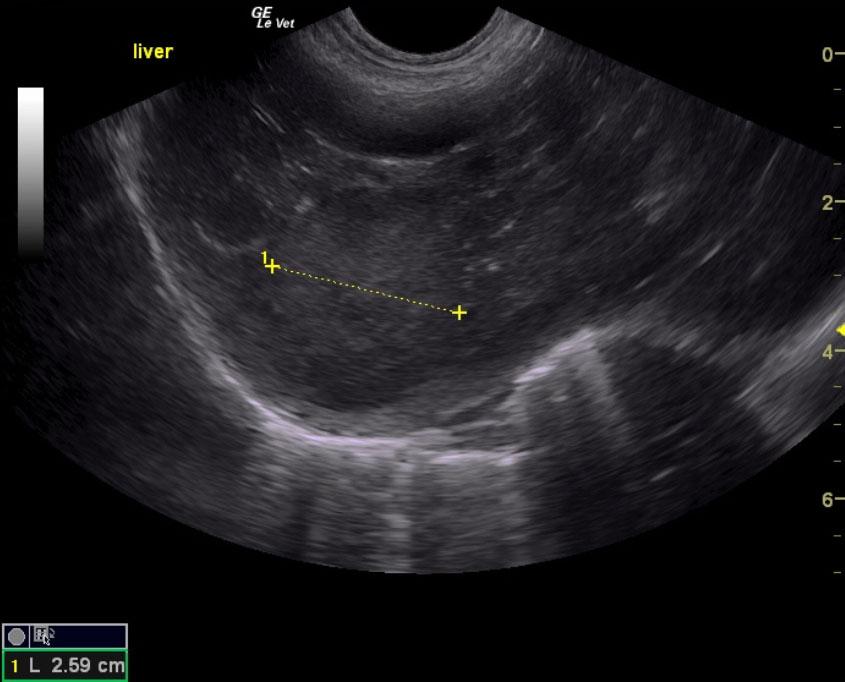
Bilateral thyroid masses. This is consistent with carcinoma. Furthermore, a hepatic metastatic lesion is suspected.

Image Interpretation

The right cervical region revealed a 5 x 4 cm lobar mass likely deriving from residual thyroid tissue. The region of the left thyroid was enlarged and created a hypoechoic mass measuring 5.2 x 1.9 cm. Regional lymphadenopathy also appeared present. The liver presented hypoechoic nodular changes in the deep right liver measuring 2.59 cm in short axis.

DX

Bilateral thyroid masses with probable liver metastasis

Outcome

No known outcome.

Clinical Differential Diagnosis

Thyroid carcinoma, other neoplasia, granuloma, abscess

Sampling

FNA of the cervical and hepatic lesions are recommended for confirmation. No sampling was done at the time of the scan.

Patient Information

Patient Name : Molly H
Gender : Female, Spayed
Species : Canine
Type of Imaging : Ultrasound
Status : Complete
Liz Wuz Here : Yes
Code : 13_00017

Clinical Signs

  • Palpable Mass

Exam Finding

  • Palpable mass

Images

rt_massrt_mass_meas_lonrt_mass_measbetter_doplt_mass_meas_longliver_nod

Clinical Signs

  • Palpable Mass