A 5-year-old neutered male ferret with alopecia was presented for evaluation of suspected adrenal disease.
A 5-year-old neutered male ferret with alopecia was presented for evaluation of suspected adrenal disease.
A 5-year-old neutered male ferret with alopecia was presented for evaluation of suspected adrenal disease.
A 5-year-old neutered male ferret with alopecia was presented for evaluation of suspected adrenal disease.
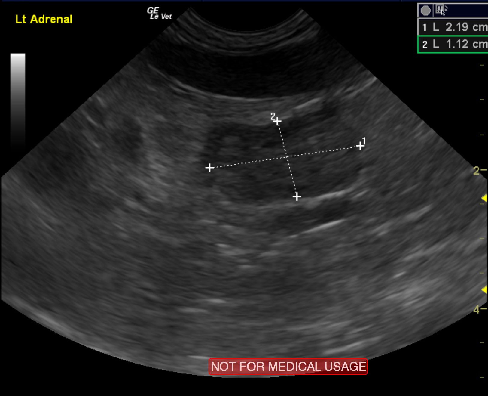
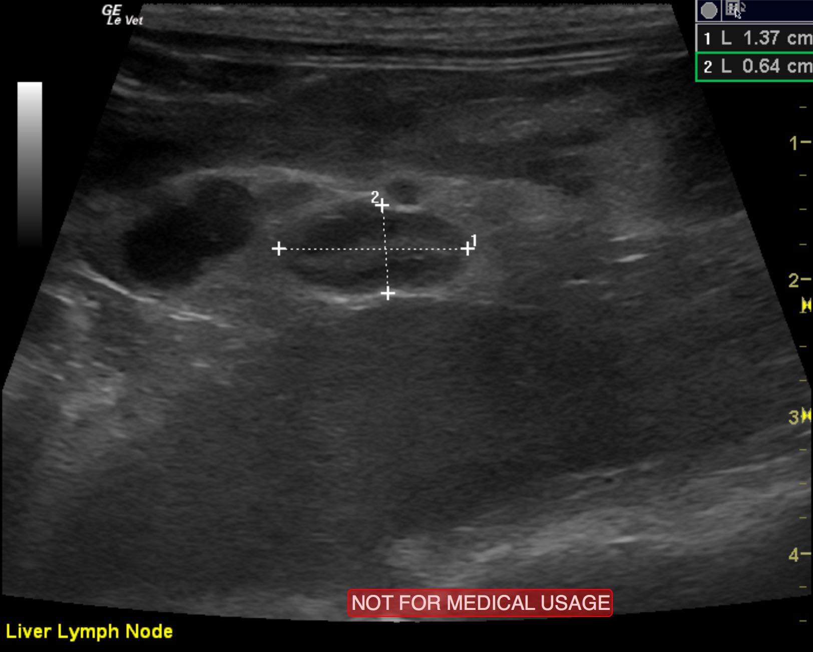
Left adrenal gland mass. Suspect carcinoma. This appears surgically resectable. Lymphadenopathy involving epigastric, splenic and portal lymph nodes. These are likely related given the contiguous pattern of associated vasculature between the spleen, gastrointestinal tract and liver in the portal system.
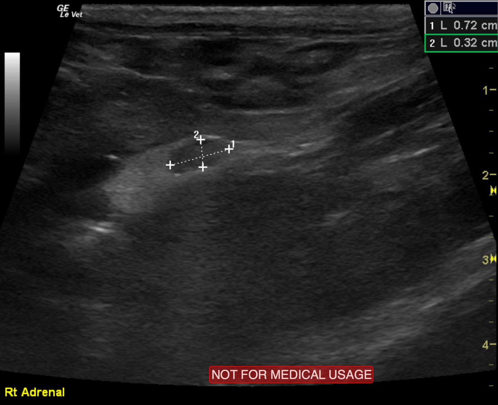
The left adrenal gland comprised a mass that measured 2.2 x 1.12 cm. Capsular expansion was noted at the caudal pole with pericapsular inflammatory pattern. It appeared significantly vascular with a trace amount of free fluid noted adjacent to it. This suggests a fairly aggressive process, but there was no evidence of vascular invasion noted. The right adrenal gland was uniform and measured 0.72 x 0.32 cm. Lymph node enlargement was noted in the epigastric region and measured 1.02 x 0.82 cm. A separate lymph node medial to the spleen was also enlarged, measuring 1 x 0.82 cm.
None
Adrenal gland disease: adenoma, carcinoma, hyperplasia.
Ultrasound guided FNA of the left adrenal gland was performed, and results were consistent with adrenocortical neoplasia. FNA of the lymph nodes were also performed, and cytology revealed heterogenous, lymphoid population, no evidence of metastasis.