An 8-year-old FS Basset Hound dog was presented for crying throughout the night and vomiting food, fluid, and mucous. On physical examination, she was found to have pink mucous membranes and a normal body temperature. She had bilateral conjunctivitis as well as a bilateral otitis externa (Malassezia). No abnormalities were noted on auscultation of her heart and lungs. The patient was treated symptomatically as an outpatient with anti-emetics and gastroprotectants. Six days later, the dog returned as she was depressed and lethargic.
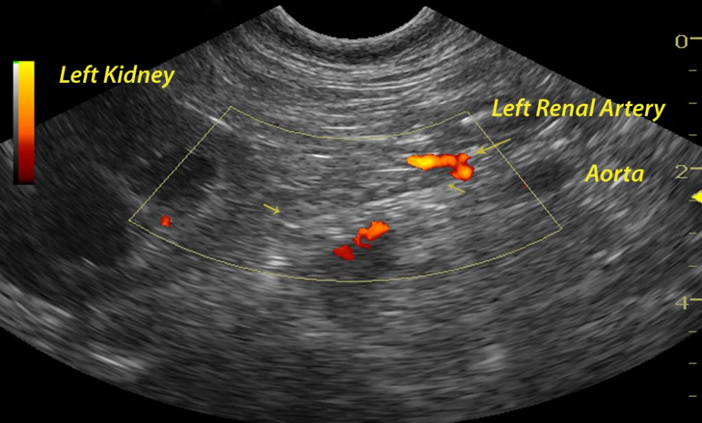
An 8-year-old FS Basset Hound dog was presented for crying throughout the night and vomiting food, fluid, and mucous. On physical examination, she was found to have pink mucous membranes and a normal body temperature. She had bilateral conjunctivitis as well as a bilateral otitis externa (Malassezia). No abnormalities were noted on auscultation of her heart and lungs. The patient was treated symptomatically as an outpatient with anti-emetics and gastroprotectants. Six days later, the dog returned as she was depressed and lethargic. She was in lateral recumbency upon presentation and she was hypothermic with pale pink, tacky and cold mucous membranes. She was admitted to the hospital for I.V. fluid therapy and blood work. The serum biochemical profile revealed elevated ALT, elevated AST, and elevated GGT, azotemia, hyperproteinemia, hyperalbuminemia, hyperglobulinemia, hyperbilirubinemia, hyperkalemia, hypercalcemia, hyperphosphatemia, hypermagnesemia, hyperamylasemia, and an elevated CK. The CBC showed an elevated hemoglobin and leukocytosis consisting of a monocytosis and eosinophilia.