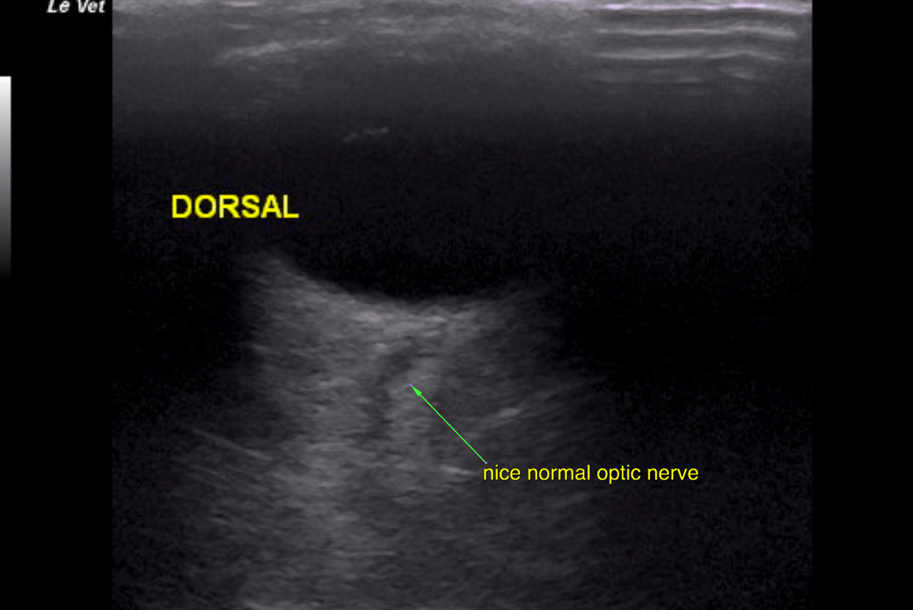
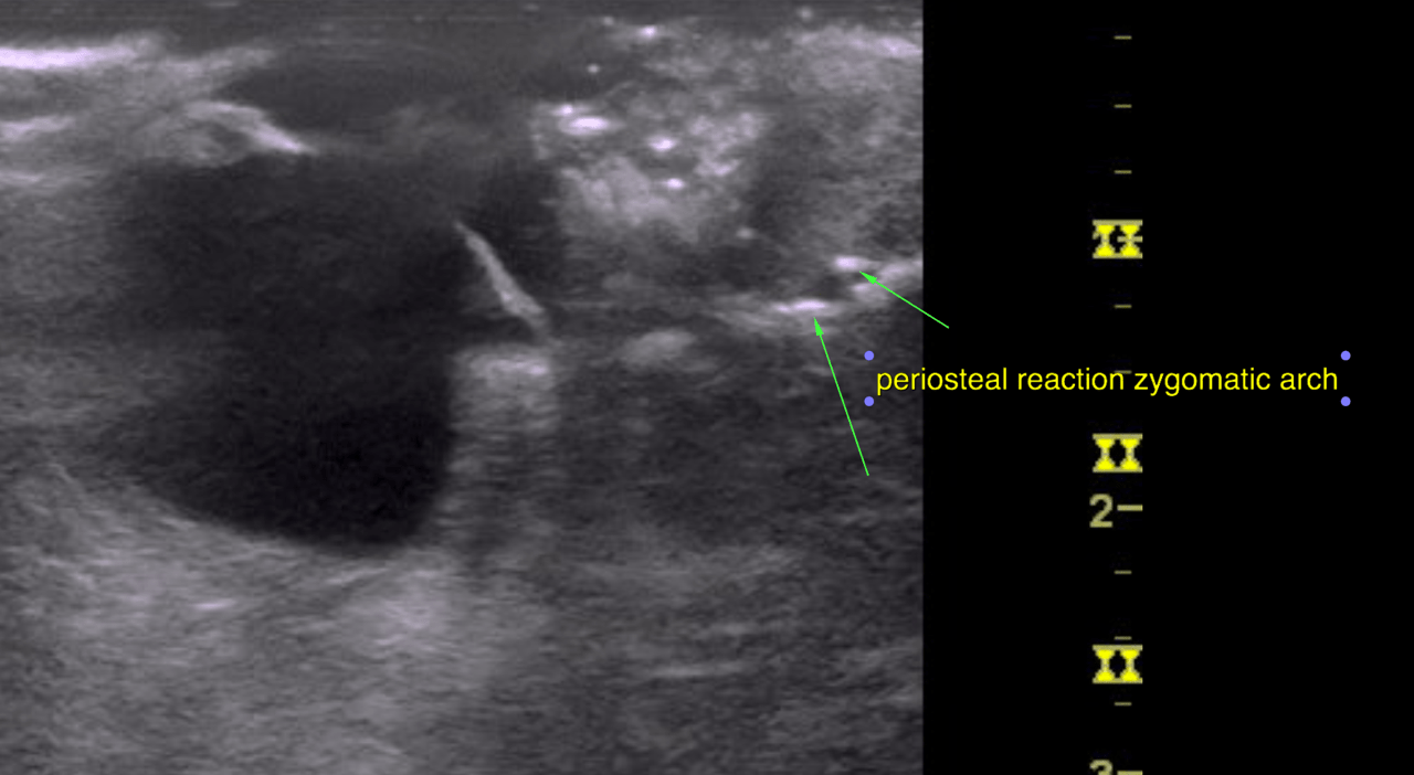
History of chronic rhinitis which is only partially responsive to steroids and no improvement with antibiotics Newly developed eye discharge, seems quieter than usual since his last Depo Medrol injection 1 month ago. Taking compounded Baytril, no other medications. Physical Exam: BARH mms pink CRT2s, Matted haircoat, BCS 5/9 – some loss of muscle mass in hind limbs, lost 1.1 lbs since last visit. EENT: Absent menace OD, retinal hemorrhage OD – seen with ophthalmoscope – limited visibility due to patient movement. Fluoro negative OU . OD periocular swelling near medial canthus.
History of chronic rhinitis which is only partially responsive to steroids and no improvement with antibiotics Newly developed eye discharge, seems quieter than usual since his last Depo Medrol injection 1 month ago. Taking compounded Baytril, no other medications. Physical Exam: BARH mms pink CRT2s, Matted haircoat, BCS 5/9 – some loss of muscle mass in hind limbs, lost 1.1 lbs since last visit. EENT: Absent menace OD, retinal hemorrhage OD – seen with ophthalmoscope – limited visibility due to patient movement. Fluoro negative OU . OD periocular swelling near medial canthus. Able to retropulse eyes. Mucopurulent nasal discharge especially on the right. Fluoro stain seen from left nostril but not the right. No arrhythmias or murmurs ausculted, referred upper airway noise, no adventitious lung sounds. BP readings averaged 215 systolic, 144 diastolic, MAP of 162 . New problems include – retinal hemorrhage, hypertension (also could be due to stress in office), periorbital swelling. Concern for mass effect, or infectious process impacting right eye.