An 8-year-old MN Shih Tzu was presented for evaluation of a heart murmur. Systolic BP was normal.
An 8-year-old MN Shih Tzu was presented for evaluation of a heart murmur. Systolic BP was normal.
Case Study
15_00082 Mikey G Mild DMVD
Sonographic Differential Diagnosis
This is a case of mild DMVD (Stage B1) causing mild mitral insufficiency.
Image Interpretation
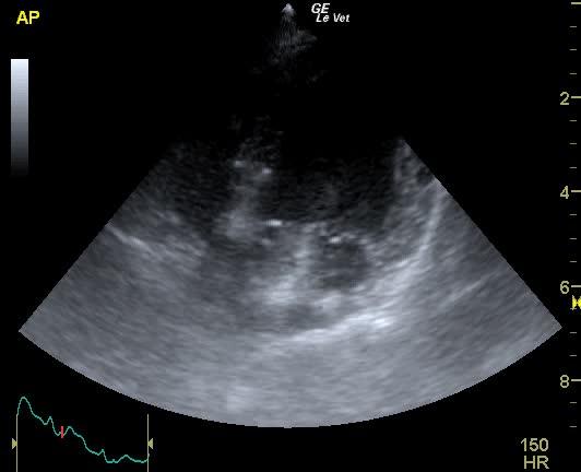
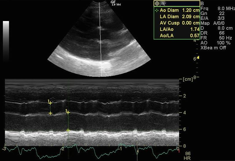
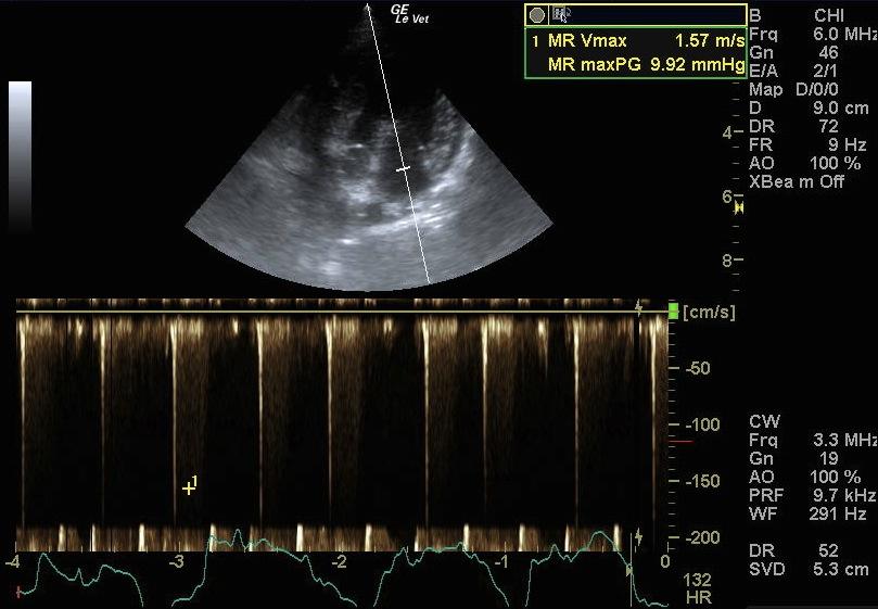
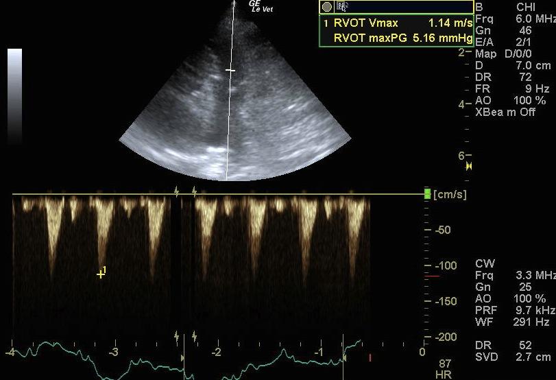
This study reveals a mildly enlarged left atrium, with a normal left ventricle. The mitral valve leaflets are mildly to moderately thickened with both leaflets slightly prolapsing into the left atrium. The mitral insufficiency noted on CDI occupies ~25% of the left atrium. Flows across the LVOT and RVOT are within normal ranges (in terms of velocity) but the flow profile across the RVOT is somehow asymmetric. This could indicate some pulmonary hypertension. However, because there is no concentric hypertrophy of the right ventricle present, the asymmetry to the RVOT flow profile should not be over-interpreted.
DX
Mild DMVD (Stage B1) causing mild mitral insufficiency.
Outcome
No therapy currently indicated. Reevaluation of the patient every 6 months is recommended.
Clinical Differential Diagnosis
Mitral/tricuspid endocardiosis, vegetative endocarditis, myocardial weakness, cardiomyopathy.
Sampling
None
Video
Patient Information
Gender :
Male, Neutered
Species :
Canine
Type of Imaging : Ultrasound
Status :
Complete
Clinical Signs
- Heart Murmur
Images
Clinical Signs
- Heart Murmur