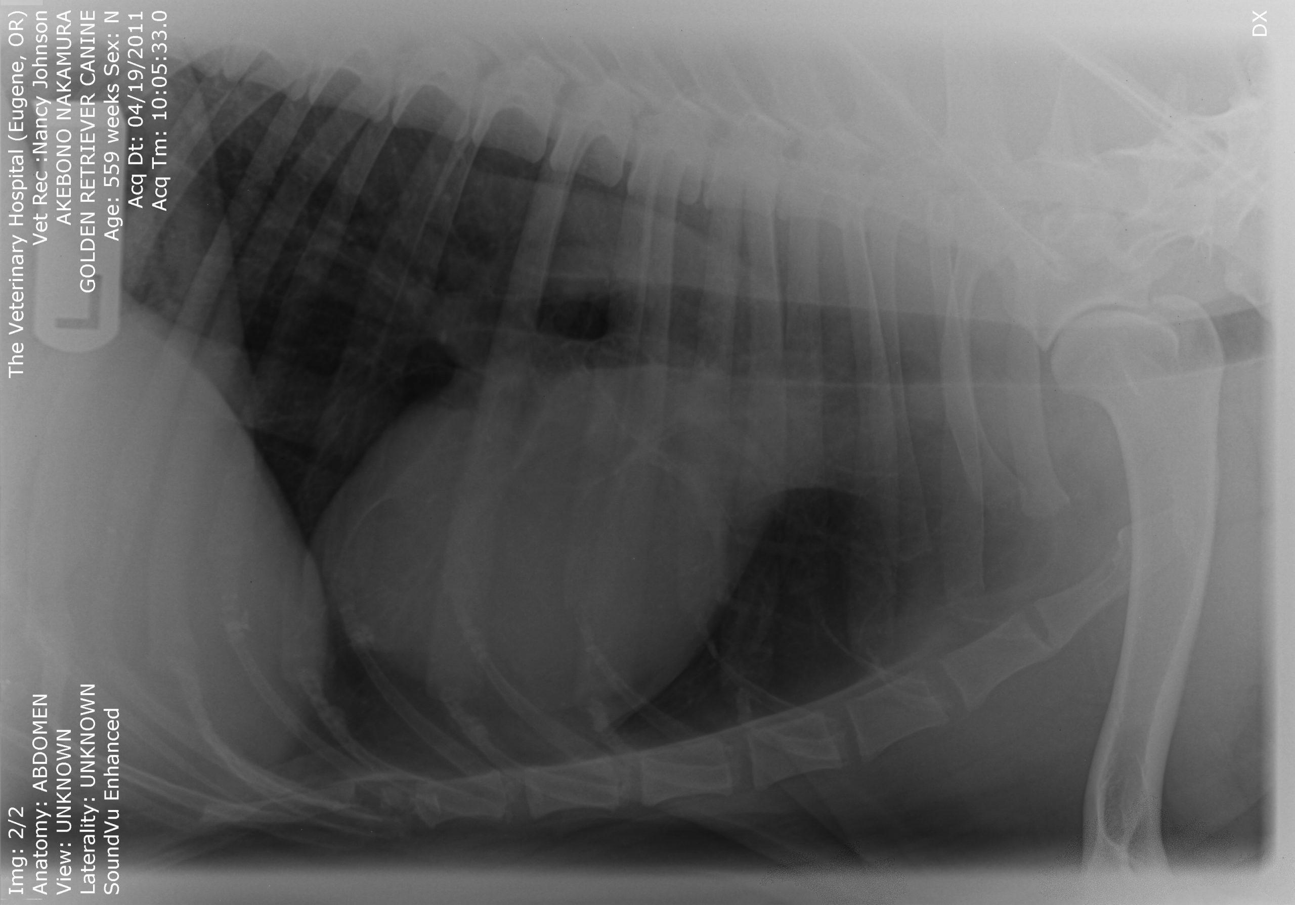
An 11-year-old MN Golden retriever was presented for evaluation due to two episodes of collapse and cyanosis. Additional history was that the patient was more lethargic than usual. Cardiac auscultation did not reveal any abnormalities and blood pressure was normal. Survey radiographs showed a mildly enlarged cardiac silhouette (VHS 11), prominent right ventricle, normal left atrial, and a normal lung pattern.
An 11-year-old MN Golden retriever was presented for evaluation due to two episodes of collapse and cyanosis. Additional history was that the patient was more lethargic than usual. Cardiac auscultation did not reveal any abnormalities and blood pressure was normal. Survey radiographs showed a mildly enlarged cardiac silhouette (VHS 11), prominent right ventricle, normal left atrial, and a normal lung pattern.
Case Study
15_00054 Akebono N Pulmonary artery masses **Nov 2011 COM**
Sonographic Differential Diagnosis
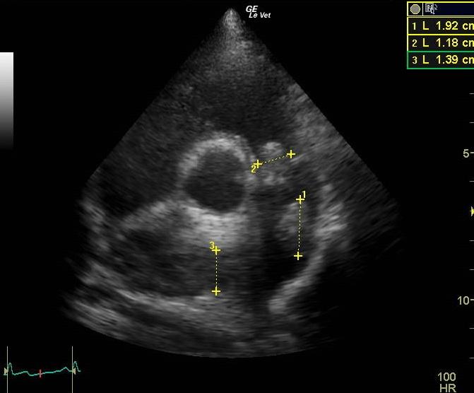
The most remarkable abnormality is the presence of a mass or masses within the pulmonary artery and attached to the pulmonic valve. All masses have the same echogenicity and cause obstruction to flow which the consequence of right ventricular hypertrophy. Even though it cannot be ruled out that these masses are thrombi (but not septic thrombi because there is no history of fever) I would think that this is neoplasia (thrombosis usually causes pulmonary hypertension). One case of pulmonary artery leiomyosarcoma is mentioned in the veterinary literature, more can be found in human literature. I would recommend administration of Plavix or aspirin in this patient.
Image Interpretation
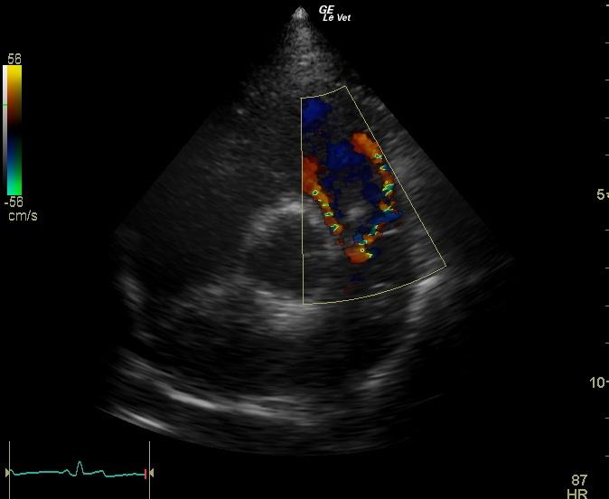
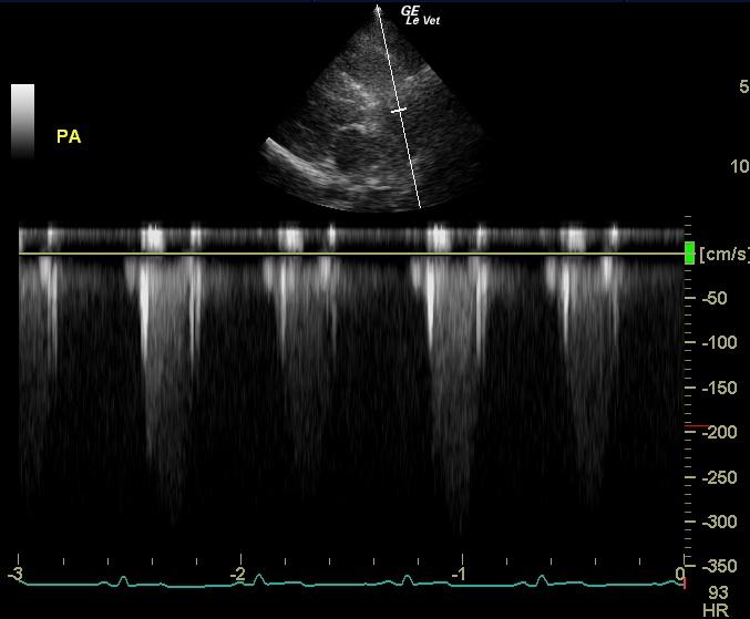
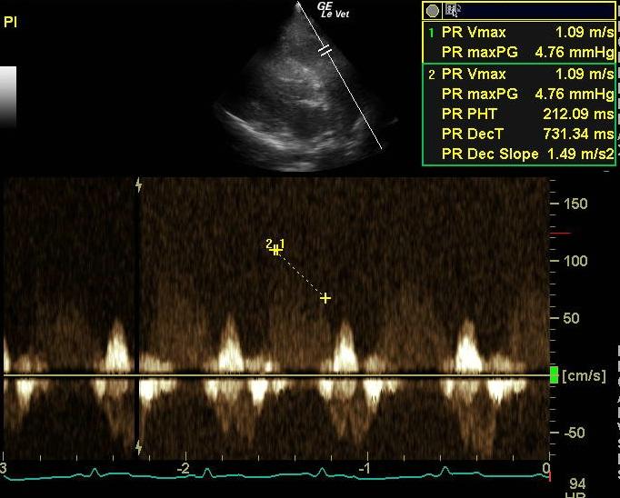
The right ventricle is mildly enlarged (hypertrophied), but no significant tricuspid valve regurgitation is noted. The right atrium displays mildly increased size. The RVOT does not show changes. There is a mass (1.18 cm) attached to and moving with the pulmonary valve. Another mass (1.92 cm) is seen along the craniodorsal border of the PA. The right pulmonary artery branch is obviously filled with another homogenous mass. On CDI it can be seen very clearly that the masses are causing obstruction to flow and seem to be somehow confluent on these clips. Flow velocity across the RVOT and PV is increased (3.2 m/s), but vmax of the pulmonary insufficiency noted is normal excluding pulmonary hypertension.
DX
Outcome
Post mortem biopsy revealed marked lung congestion, right ventricular hypertrophy, and grade 3 fibrosarcoma of the pulmonary valve with attached thrombus.
Comments
The owners opted for euthanasia and necropsy.
Clinical Differential Diagnosis
Pericardial effusion. Cardiomyopathy – hypertrophic, dilated. Myocarditis. Cardiac neoplasia. Pulmonic thromboembolic disease. Metabolic disease – anemia, methemoglobinemia.
Sampling
None
Video
Patient Information
Clinical Signs
- Collapse
- Cyanosis
- Lethargy
Images
Clinical Signs
- Collapse
- Cyanosis
- Lethargy