A 5-year-old intact male Beagle with pneumonia that was responding to antibiotic therapy was presented for evaluation of a heart murmur.
A 5-year-old intact male Beagle with pneumonia that was responding to antibiotic therapy was presented for evaluation of a heart murmur.
1.) Patent Ductus Arteriosus – continuous L to R flow
2.) Severe LVE and myocardial failure
3.) Pneumonia – recommend review of serial radiographs
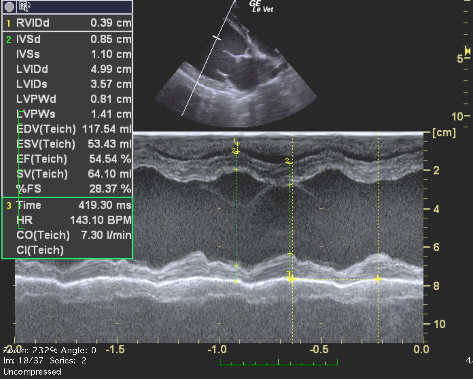
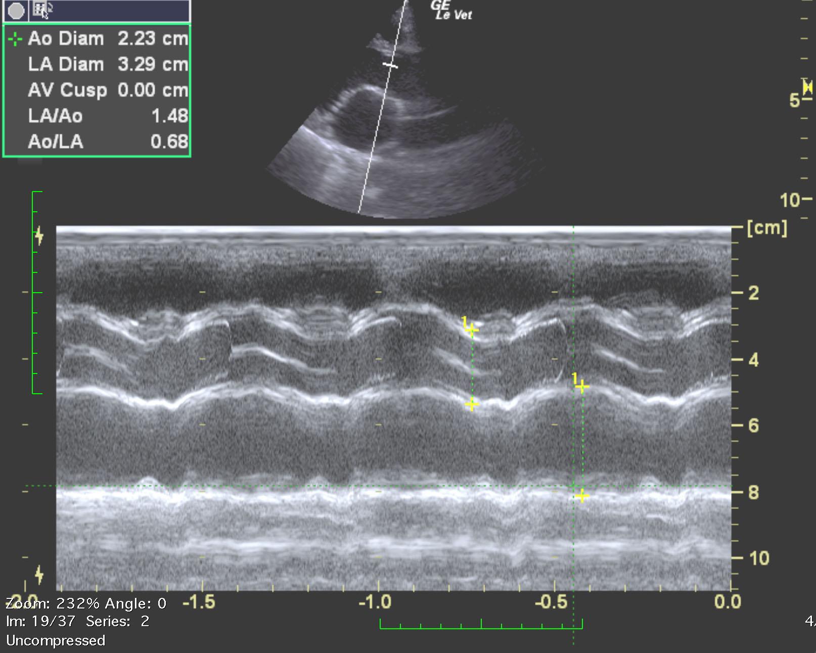
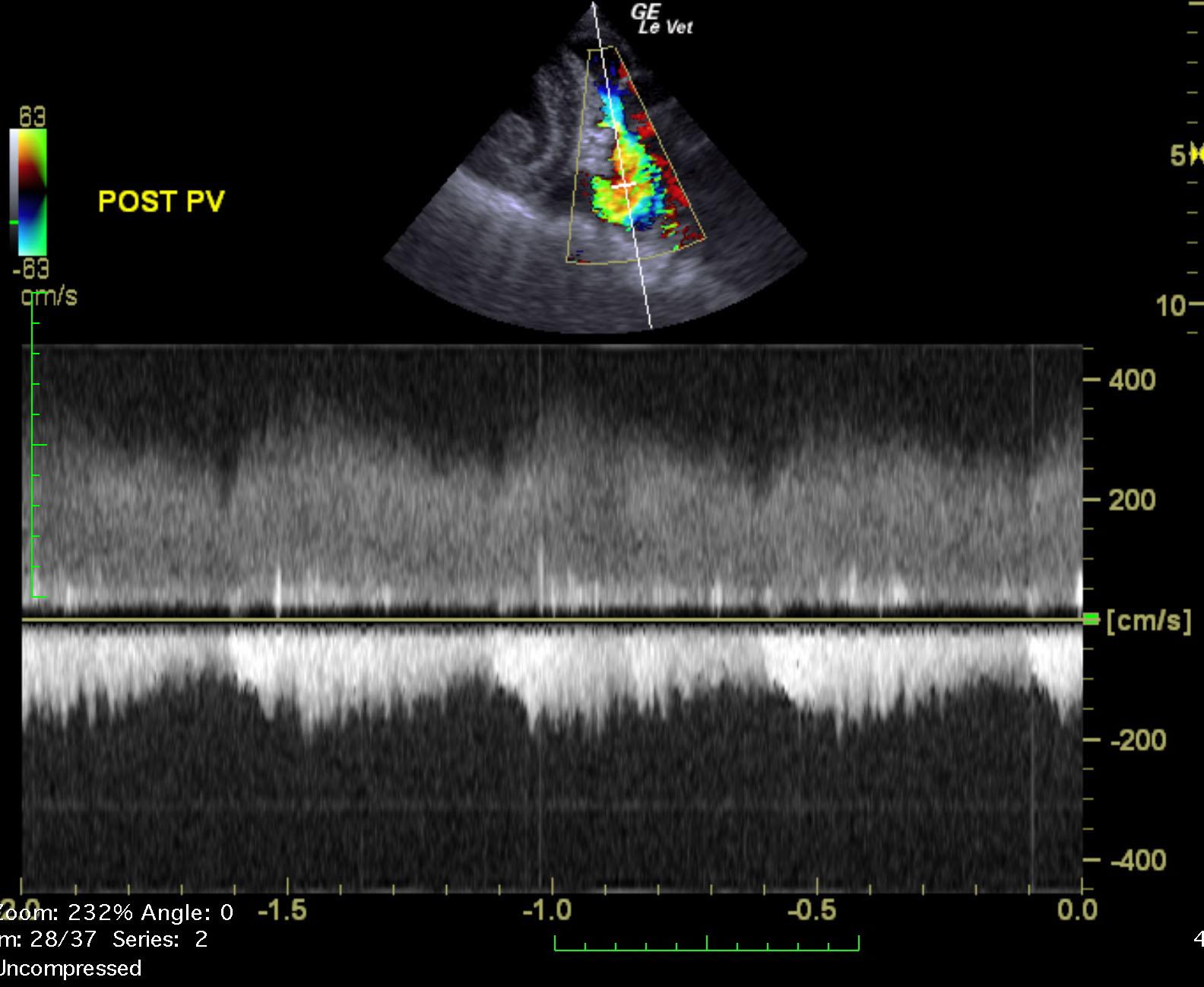
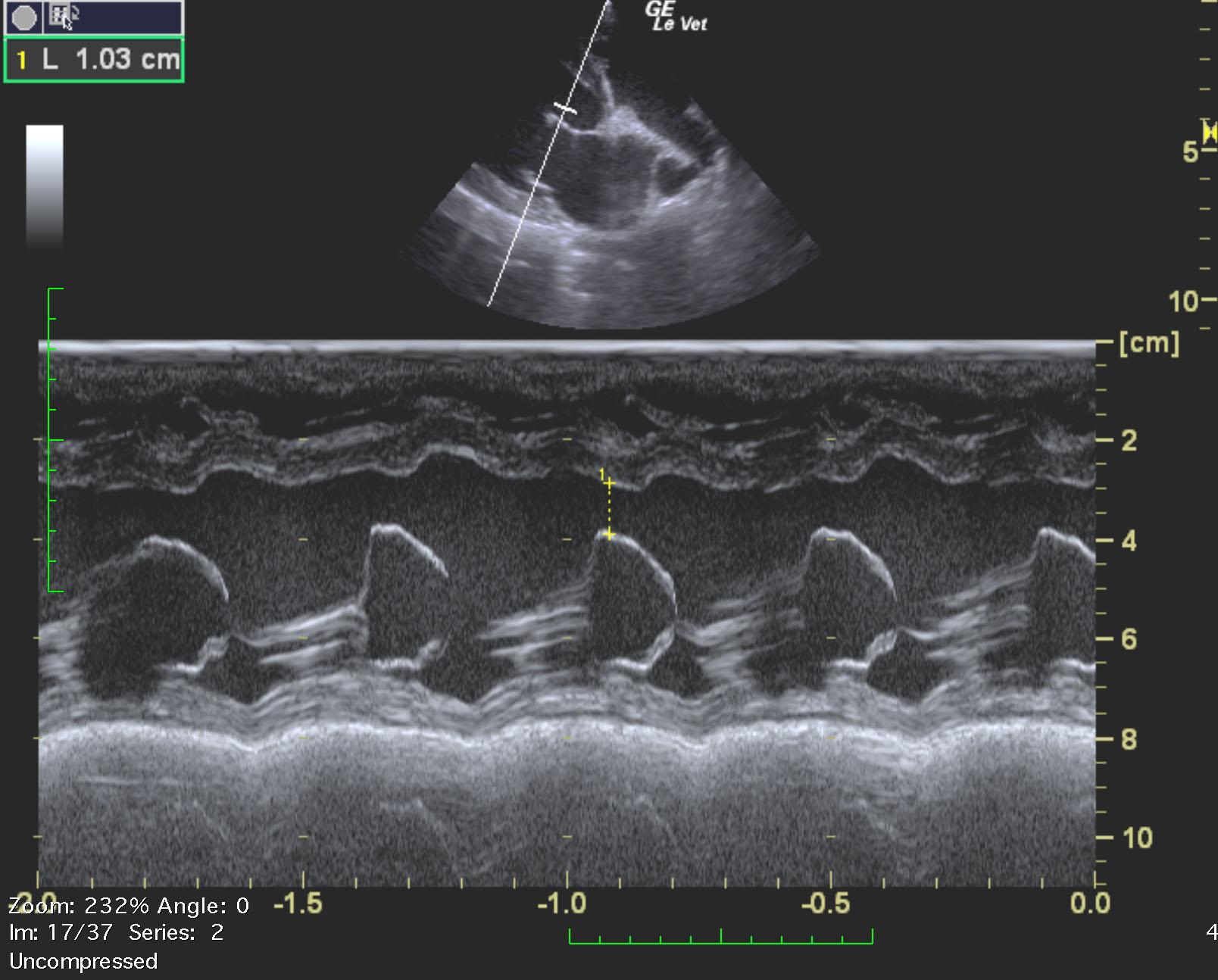
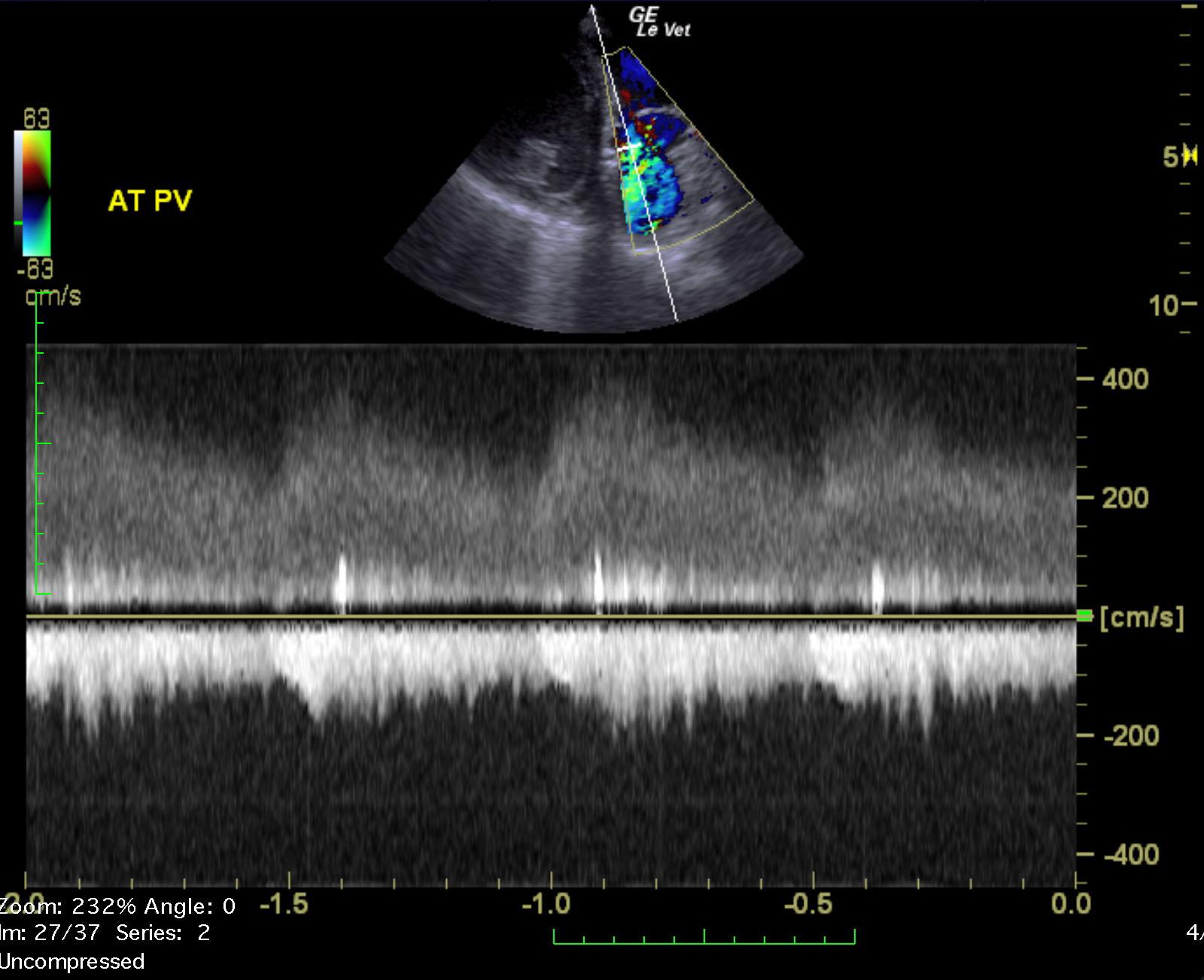
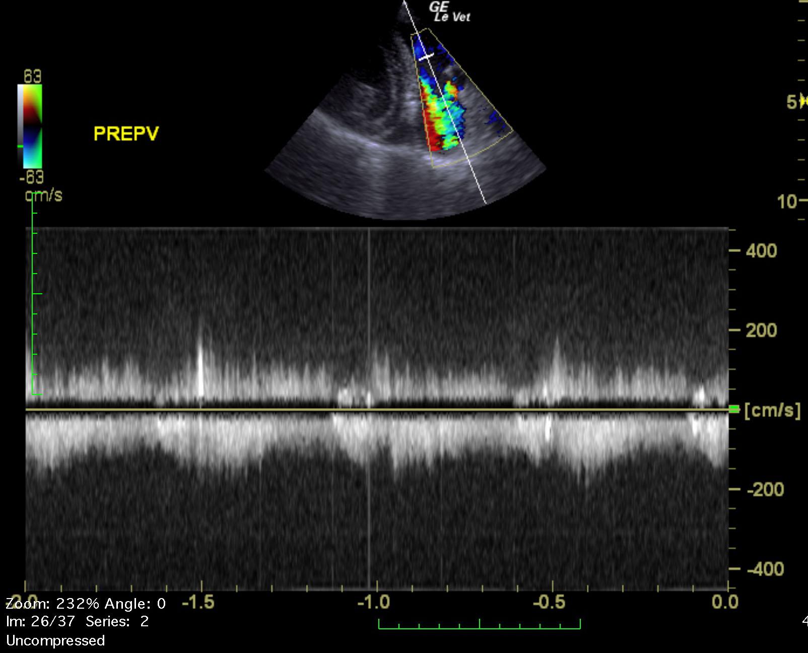
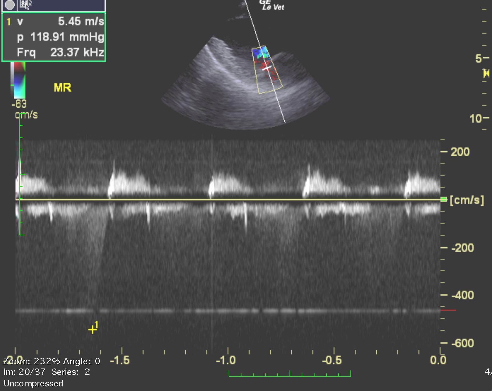
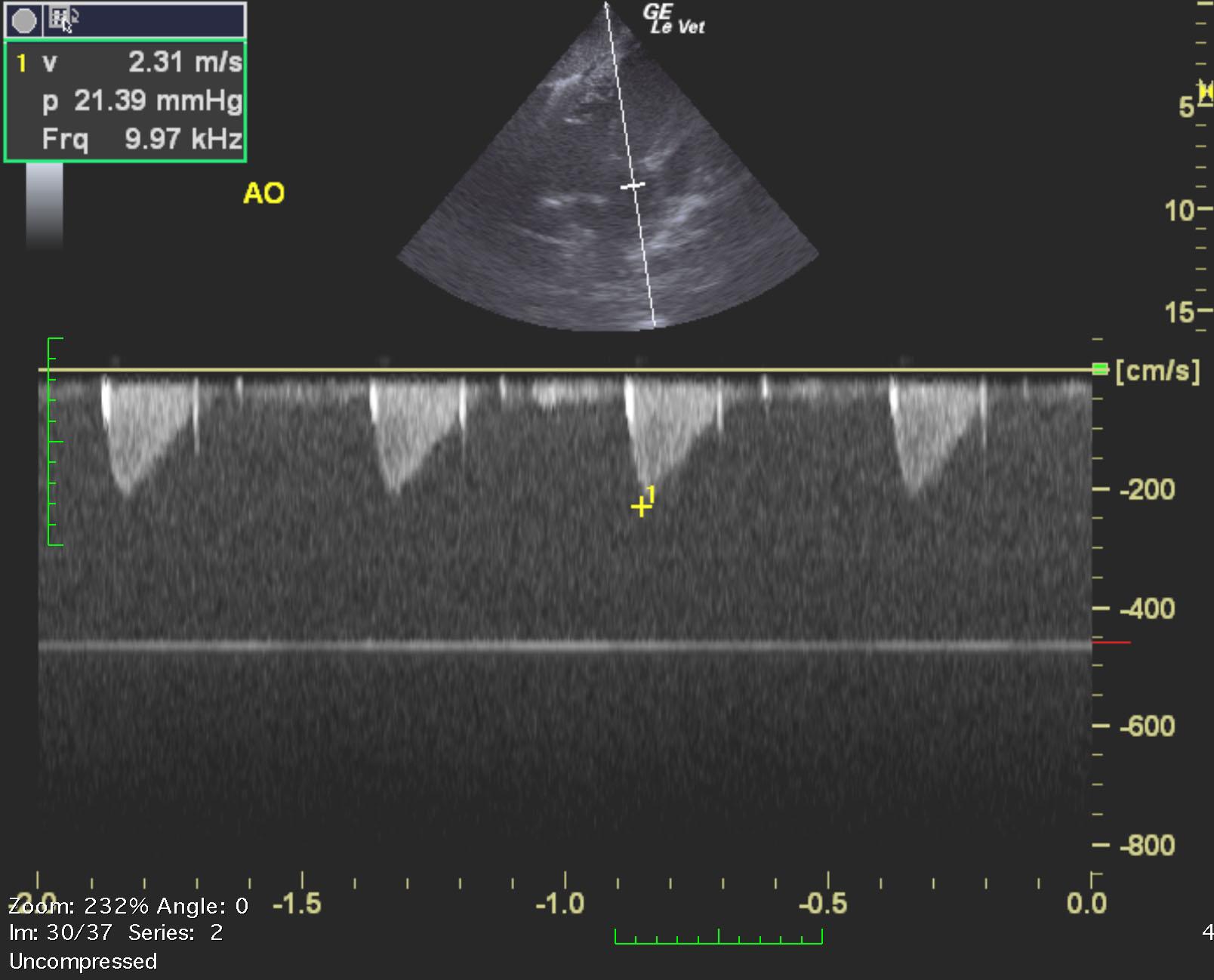
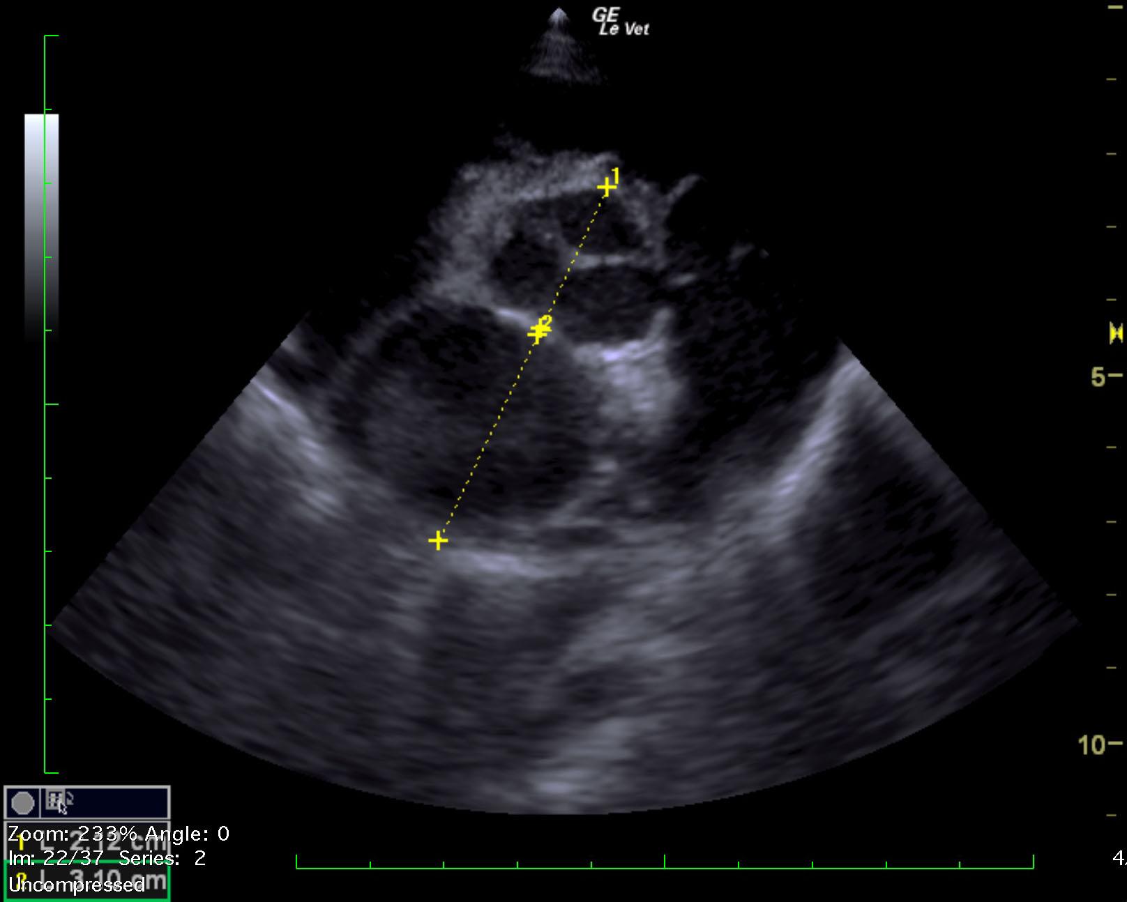
The left ventricle measures severely increased in size with notable myocardial failure. The left atrium is mildly enlarged (estimated ~La:Ao 1.50). The visible right heart is normal in size. Color Doppler identifies moderate MR (peak velocity 5.45m/s). The MPA appears focally dilated just distal to the PV. Continuous L-to-R flow can be visualized entering the branching MPA via color and spectral Doppler (peak systolic velocity ~4m/s, end diastolic velocity ~3m/s). The PDA vessel itself is not visualized. There is mildly elevated flow across the LVOT/AV (LVOT 2.31m/s). The branching pulmonary arteries are enlarged. No effusions or masses visualized. IVDd – 0.85cm LVIDd – 4.99cm LVFWd – 0.81cm IVDs – 1.10cm LVIDs – 3.57cm LVFWs – 1.41cm FS – 28.4%
None
Vegetative endocarditis, myocarditis, congenital cardiac defect (PDA, pulmonic stenosis, atrial/septal defect)
None