A 14-year-old NM German shepherd was presented for evaluation of a right neck mass.
Case Study
13-00027 Knox C Thyroid carcinoma ——–NEED SB———-
Sonographic Differential Diagnosis
Mineralizing right thyroid mass with regional lymphadenopathy.
FNA in the mineralized area and non mineralized area were performed without complication.
Image Interpretation
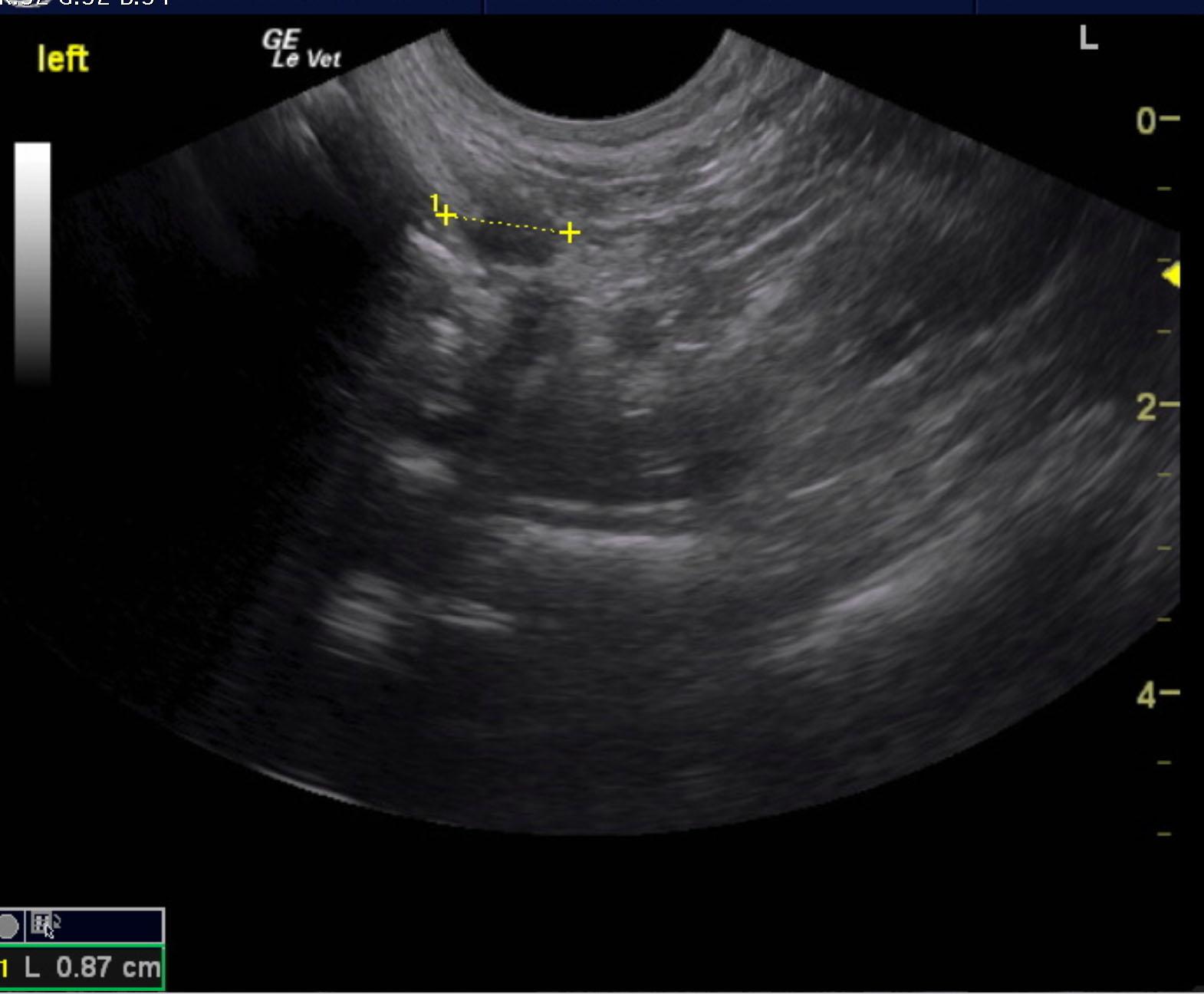
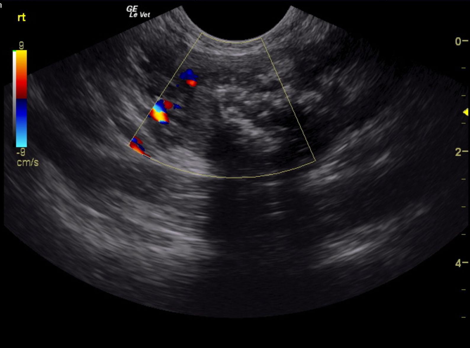
The left thyroid lobe appeared uniform and measured 0.87 cm. The left thyroid lobe was mildly hypoechoic to the surrounding fat. The right thyroid lobe revealed a mineralizing mass that measured 2.0 cm. Significant color flow Doppler signals were noted in the right thyroid region. Regional lymph nodes appeared enlarged. Clean resection is unlikely.
Outcome
None
Clinical Differential Diagnosis
Neoplasia/abscess/cyst/granuloma – lymph node, thyroid, muscle
Hematoma
Abscess
Sampling
???
Video
Patient Information
Gender :
Male, Neutered
Species :
Canine
Type of Imaging : Ultrasound
Clinical Signs
- Palpable Mass
Images
Clinical Signs
- Palpable Mass