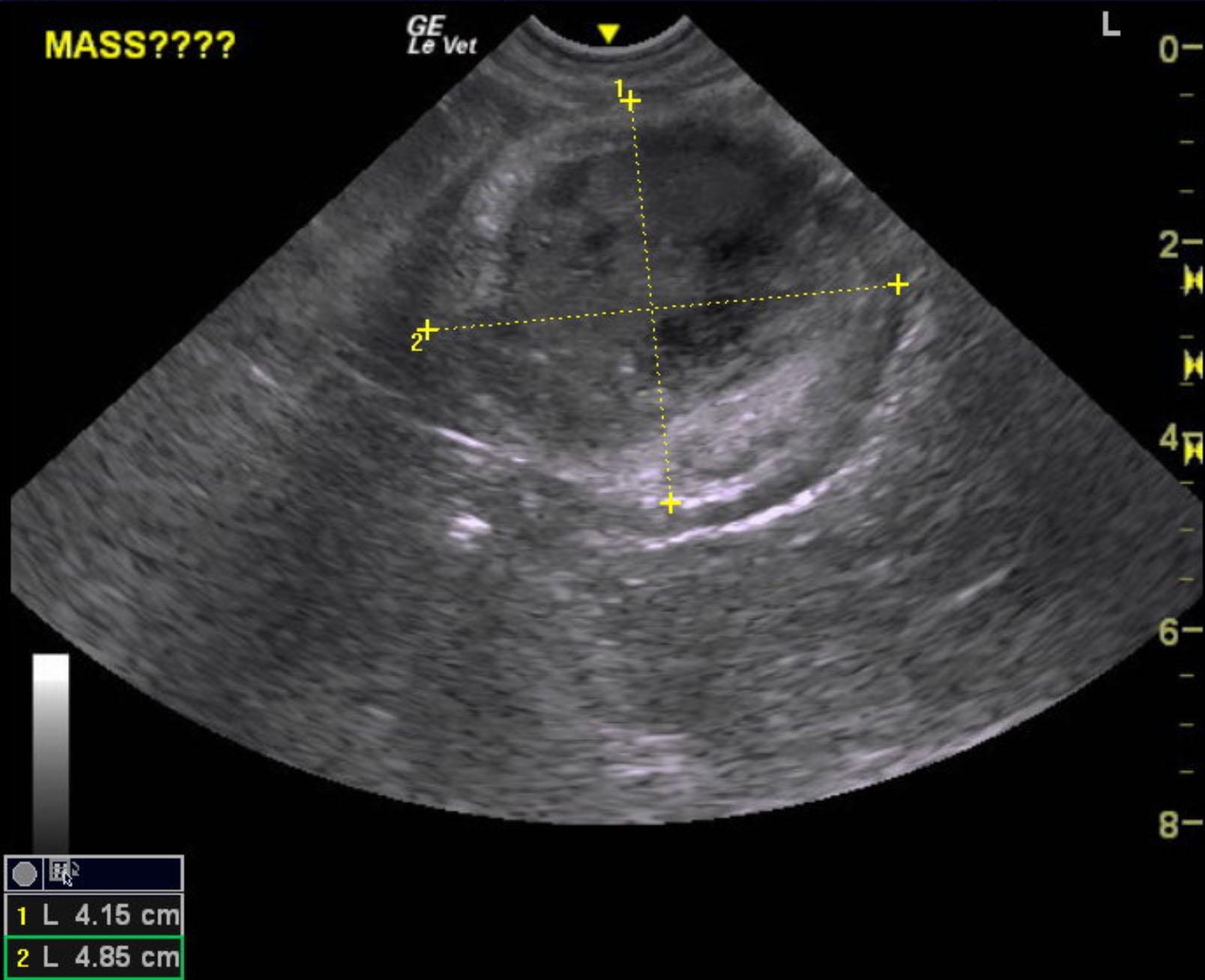
A 14-year-old SF Kooikerhendie was presented for evaluation of a mid-caudal abdominal mass and vestibular syndrome. CBC and serum biochemistry were both within reference range.
Sonographic Differential Diagnosis
Age related abdomen, kidneys, spleen and liver.
Omental abscess type mass.
Image Interpretation
A mixed echogenic mass was noted in the mid cranial abdomen and measured 4.85 x 4.15 cm with a hyperechoic, granuloma type border. This mass appears to be free within the omentum and is most consistent with an omental abscess. Granulomatous bed was noted. Recommend exploratory surgery with removal along with culture and biopsy. No significant color flow was noted in this patient only color flow artifact on power Doppler. Therefore, this mass is largely avascular.
DX
Omental abscess
Outcome
None
Clinical Differential Diagnosis
Mass – neoplasia/cyst/abscess/granuloma of liver, spleen, kidney, mesentery, lymph node
Hydronephrosis
Vestibular syndrome – idiopathic, vascular accident, neoplasia