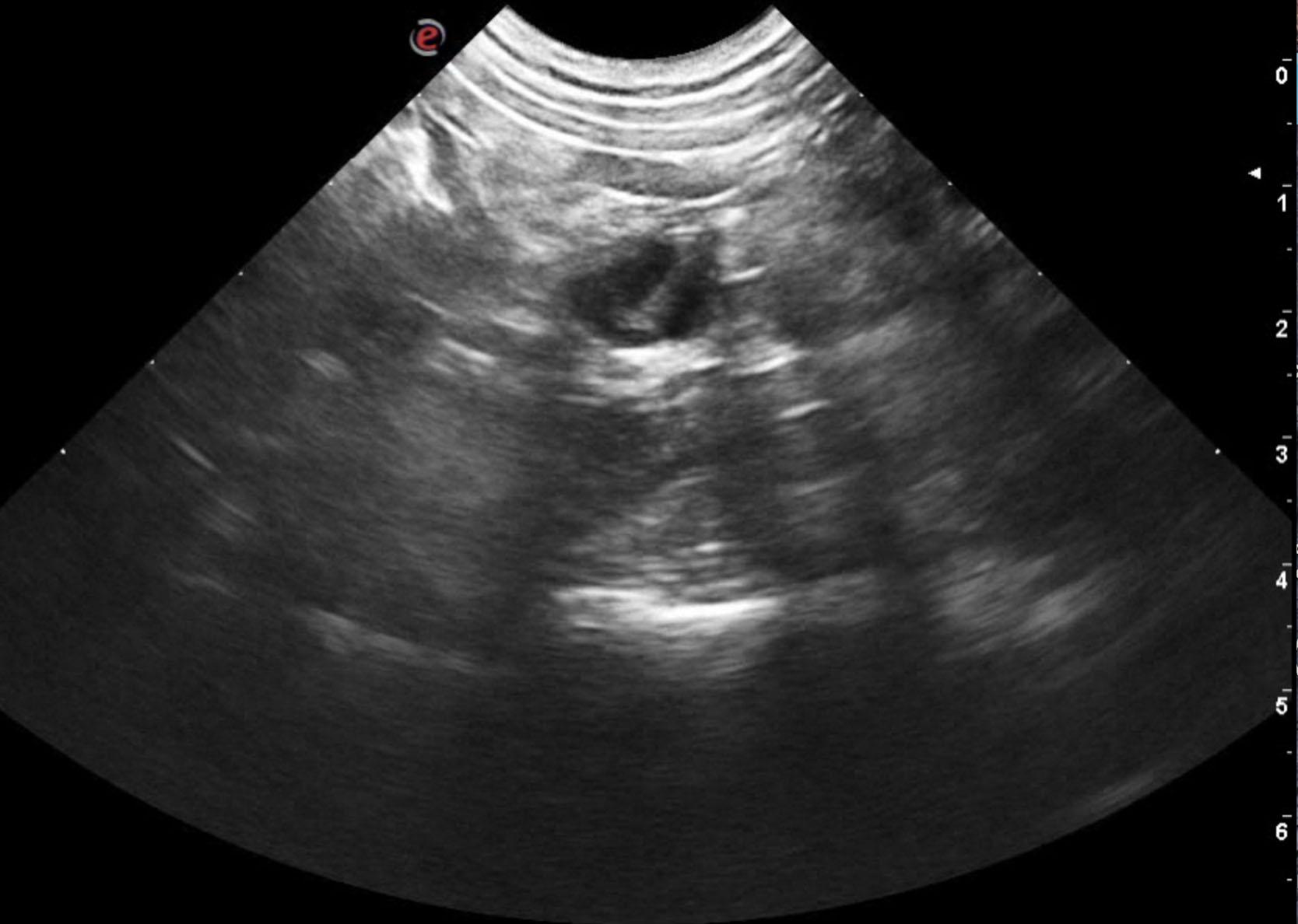
A 2-year-old SF Pug/Boston mix was presented for evaluation of signs of estrus. On physical examination, the vulva was swollen and there was a red-tinged discharge.
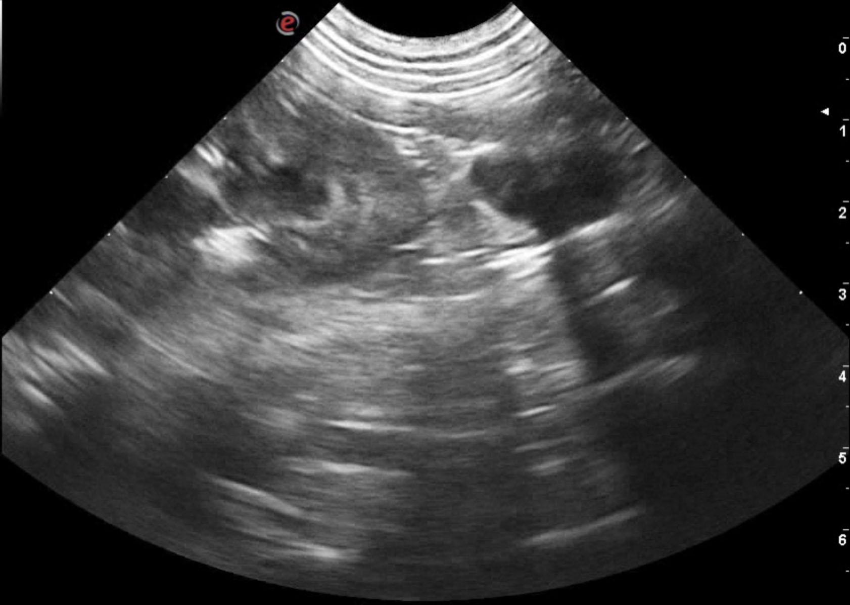
A 2-year-old SF Pug/Boston mix was presented for evaluation of signs of estrus. On physical examination, the vulva was swollen and there was a red-tinged discharge.