09_00162 Suzie A Chronic metritis

Case Study

09_00162 Suzie A Chronic metritis

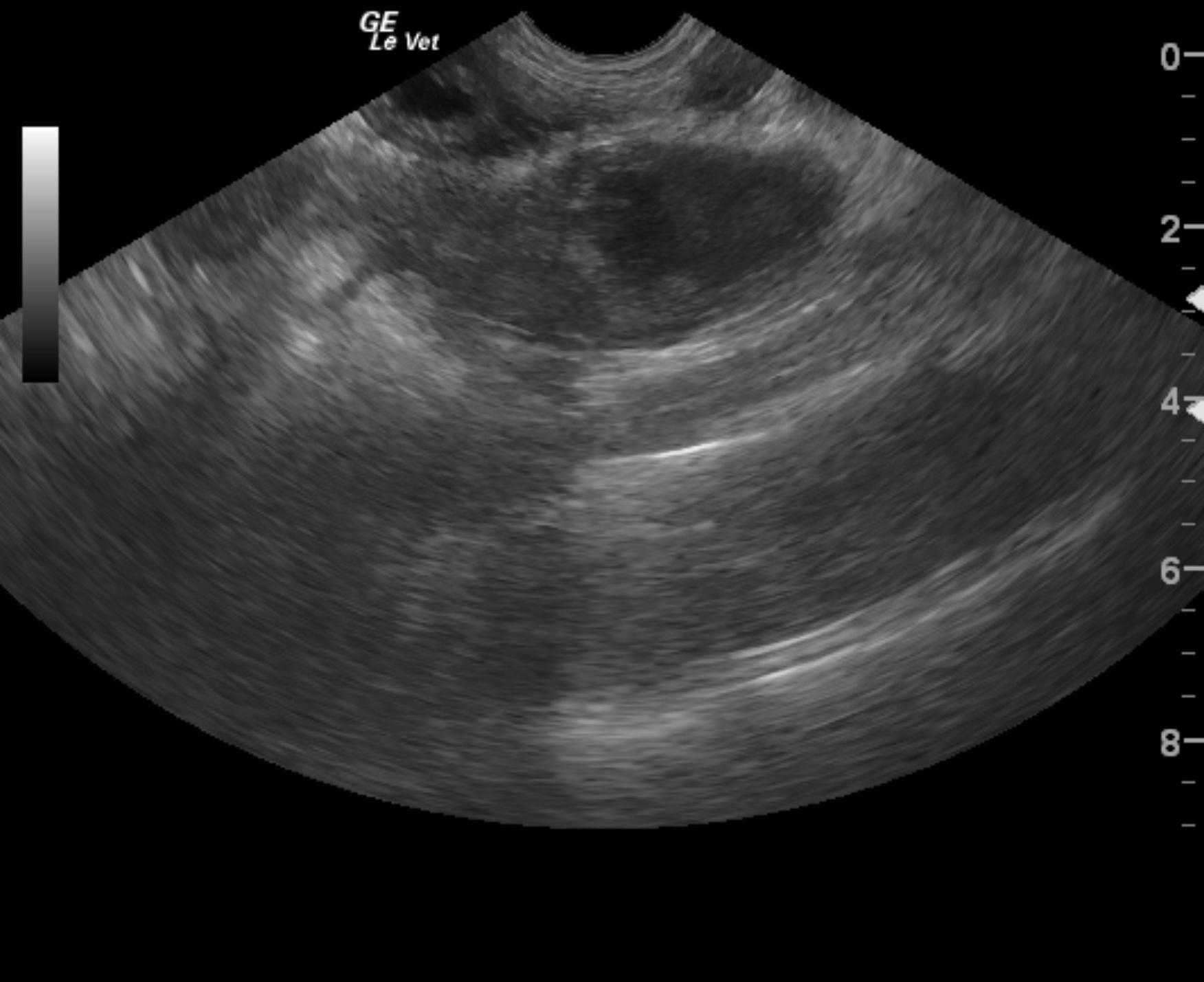
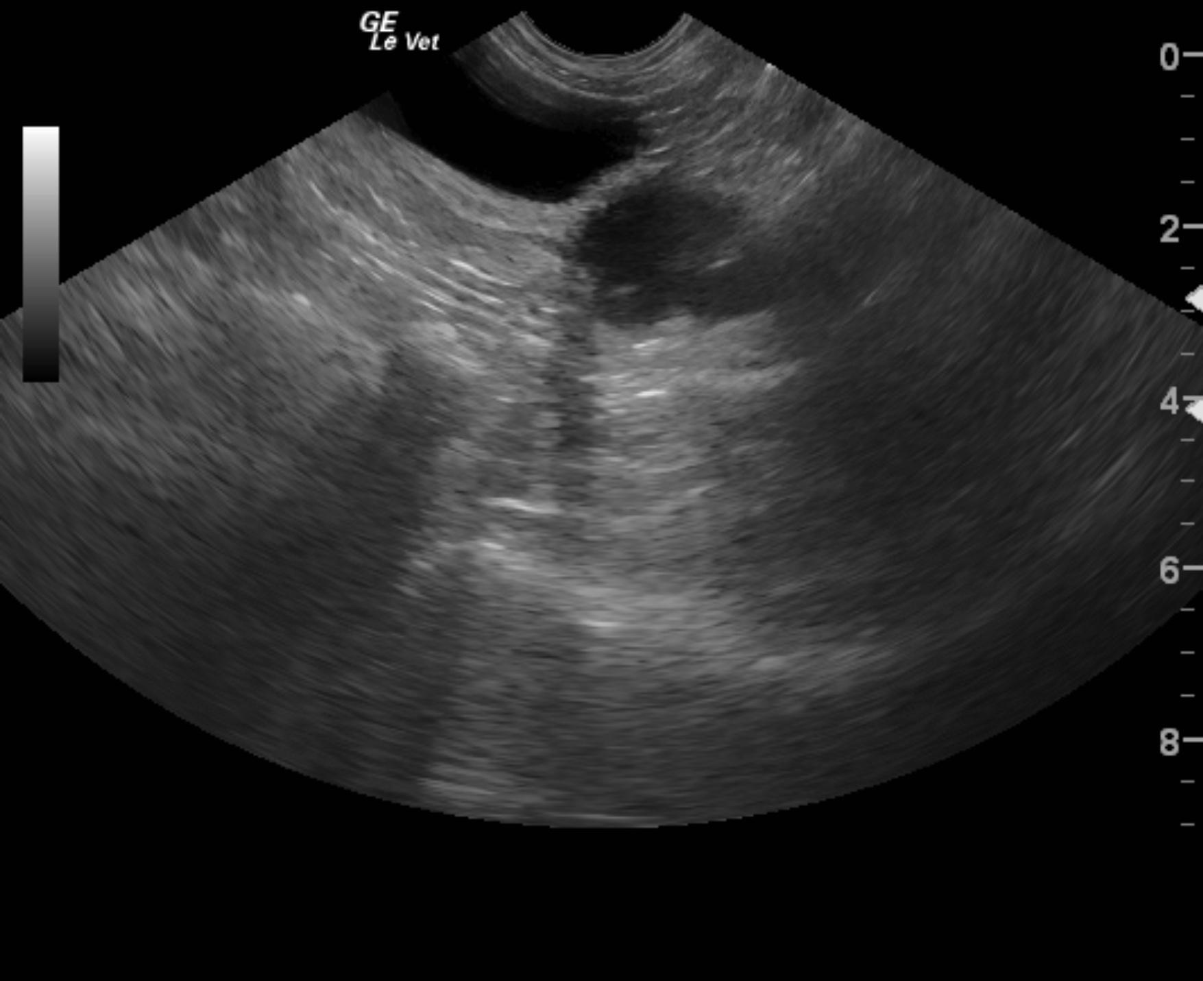
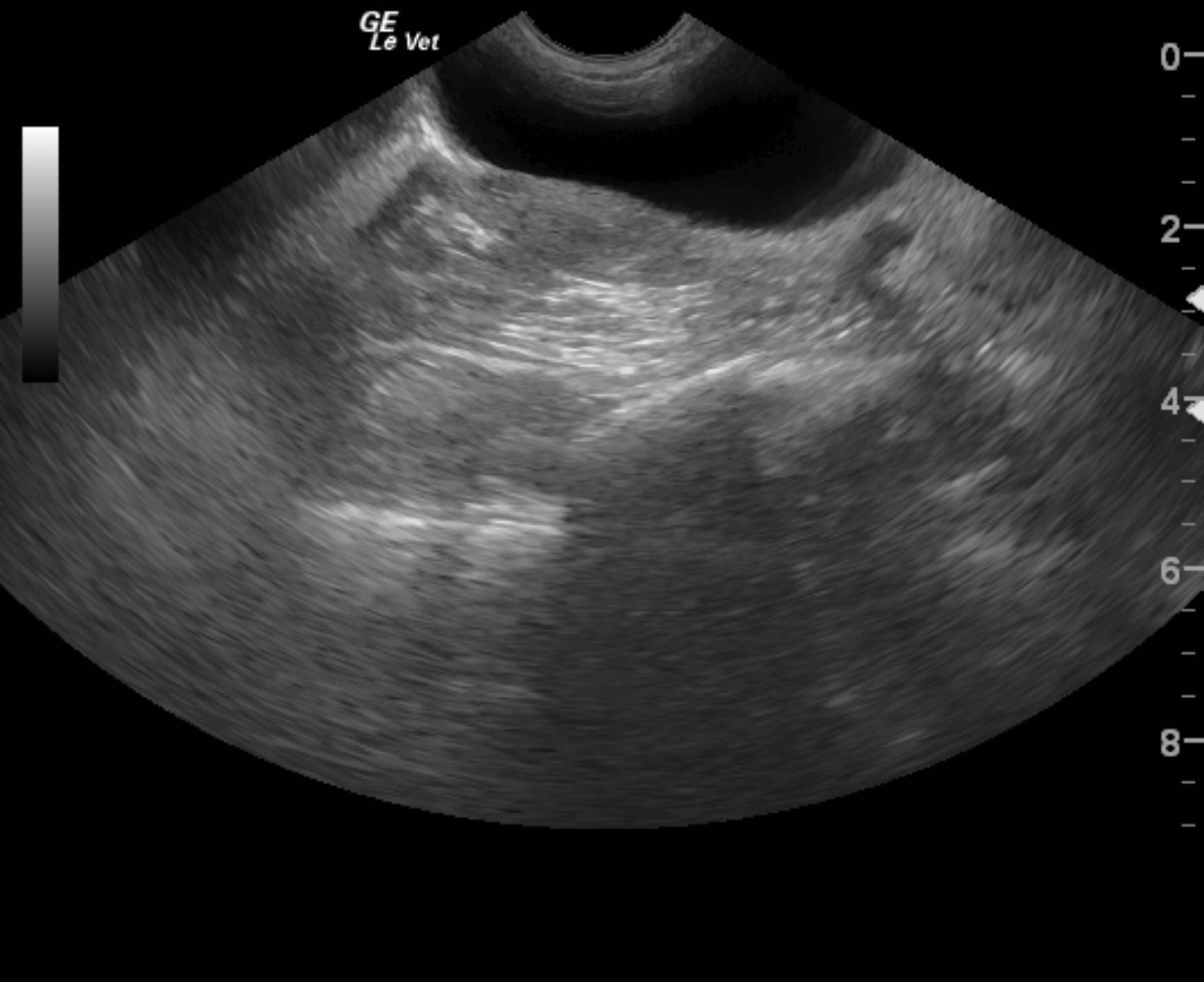
A 3-year-old female Yorkshire terrier was with a history of vaginal discharge over a few days was presented for evaluation of vomiting, diarrhea and trembling. Vaginal discharge was presented on physical examination. CBC showed mild anemia, neutrophilia, and monocytosis. Serum biochemistry was within reference range.


Sonographic Differential Diagnosis

Chronic metritis, pyometra. Mild potential for underlying neoplasia. Some of the echogenic luminal structures could technically represent mass or resorbing fetuses.

Image Interpretation

The uterus in this patient was mildly thickened measuring 1 cm in width with echogenic debris. A cystic structure was noted at the base of the uterus measuring 2 cm suggestive for fluid accumulation. Both horns of the uterus were thickened with dilated lumen and echogenic fat surrounding the uterine horns suggestive for transmural inflammation.

DX

Chronic metritis

Outcome

None

Clinical Differential Diagnosis

Metritis, cystic endometrial hyperplasia, pyometria

Sampling

None

Patient Information

Gender : Female, Intact
Species : Canine
Type of Imaging : Ultrasound
Status : Complete

Clinical Signs

  • Diarrhea
  • Shaking
  • Vaginal discharge
  • Vomiting

Exam Finding

  • Vaginal discharge

Images

almanzar_suzie_metritis_pyometraalmanzar_suzie_metritis_pyometra_2almanzar_suzie_metritis_pyometra_3

CBC

  • Monocytes, High
  • Neutrophils, High
  • RBC, Low

Clinical Signs

  • Diarrhea
  • Shaking
  • Vaginal discharge
  • Vomiting
Skip to content