A 9-year-old NM canine mix was presented for evaluation of hematuria, stranguria, and tenesmus. Urinalysis showed normal SG, hematuria, proteinuria, and leukosuria.
A 9-year-old NM canine mix was presented for evaluation of hematuria, stranguria, and tenesmus. Urinalysis showed normal SG, hematuria, proteinuria, and leukosuria.
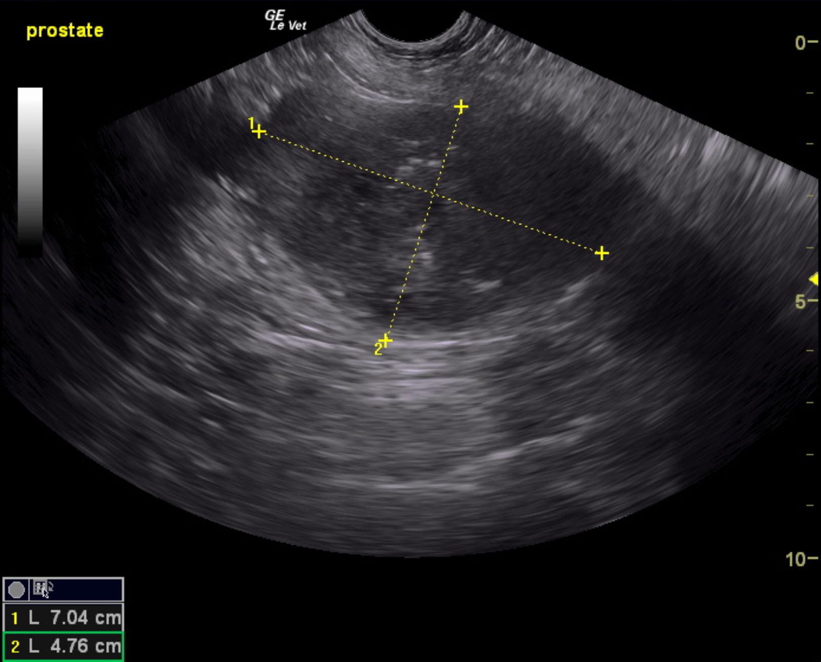
Prostatic mass.
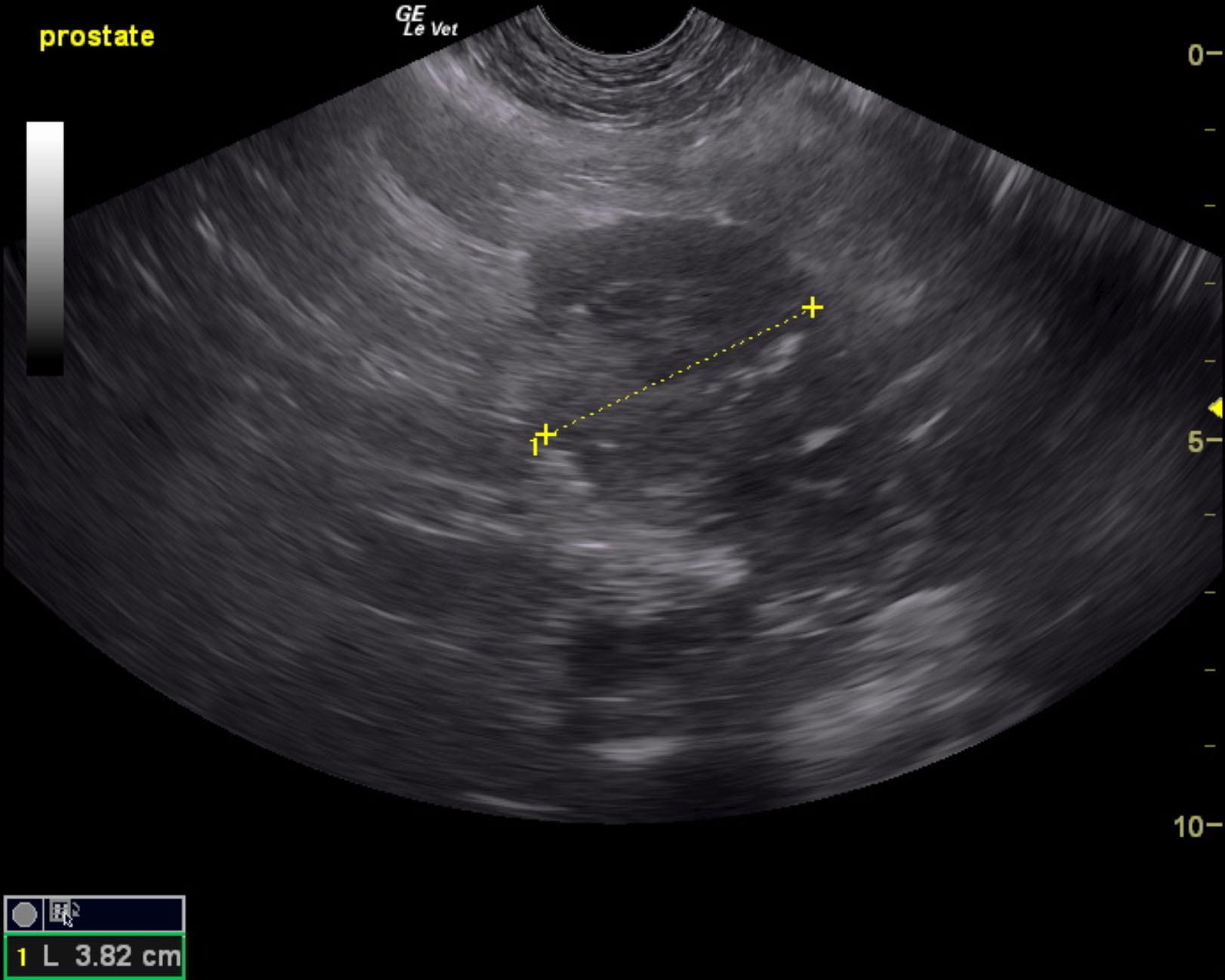
Regional inflammation.
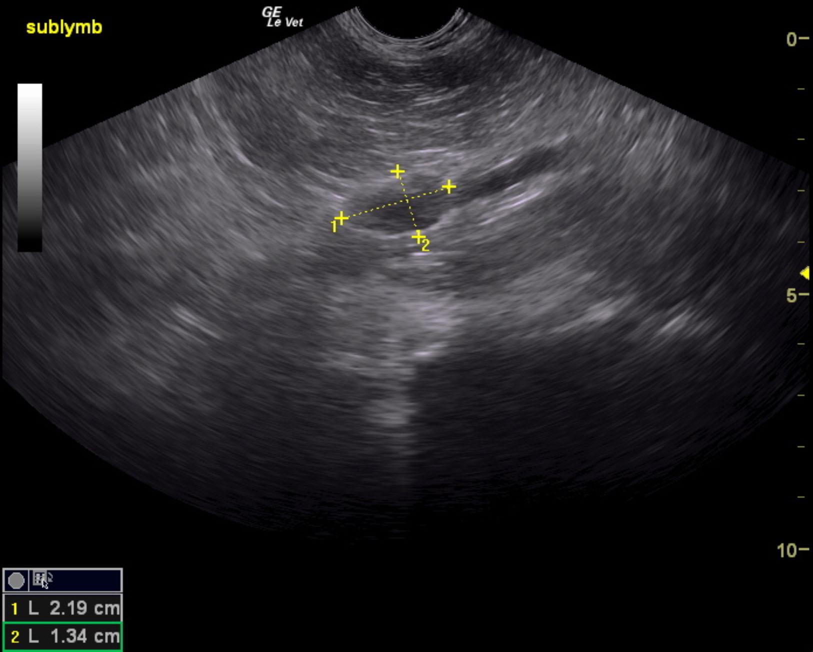
Irregular iliac lymph node. This is suggestive for early metastatic disease. Ultrasound-guided FNA would be recommended for confirmation.
The prostate in this patient was enlarged and mineralized. The prostate measured 7.04 x 4.76 cm with pericapsular inflammatory pattern. Microcystic changes were noted within the prostate.
Lymph node enlarged measuring 2.19 x 1.34 cm. The lymph node was hypoechoic and irregular with loss of length to width ratio.
None
Prostate – neoplasia, abscess, prostatitis
Bladder – neoplasia, urolith
None