A 7-year-old NM Beagle was presented for evaluation of urinating in house and tenesmus. Prostate mass was palpated on rectal. Urinalysis showed normal SG, hematuria, proteinuria, leukosuria, and epithelial cells.
A 7-year-old NM Beagle was presented for evaluation of urinating in house and tenesmus. Prostate mass was palpated on rectal. Urinalysis showed normal SG, hematuria, proteinuria, leukosuria, and epithelial cells.
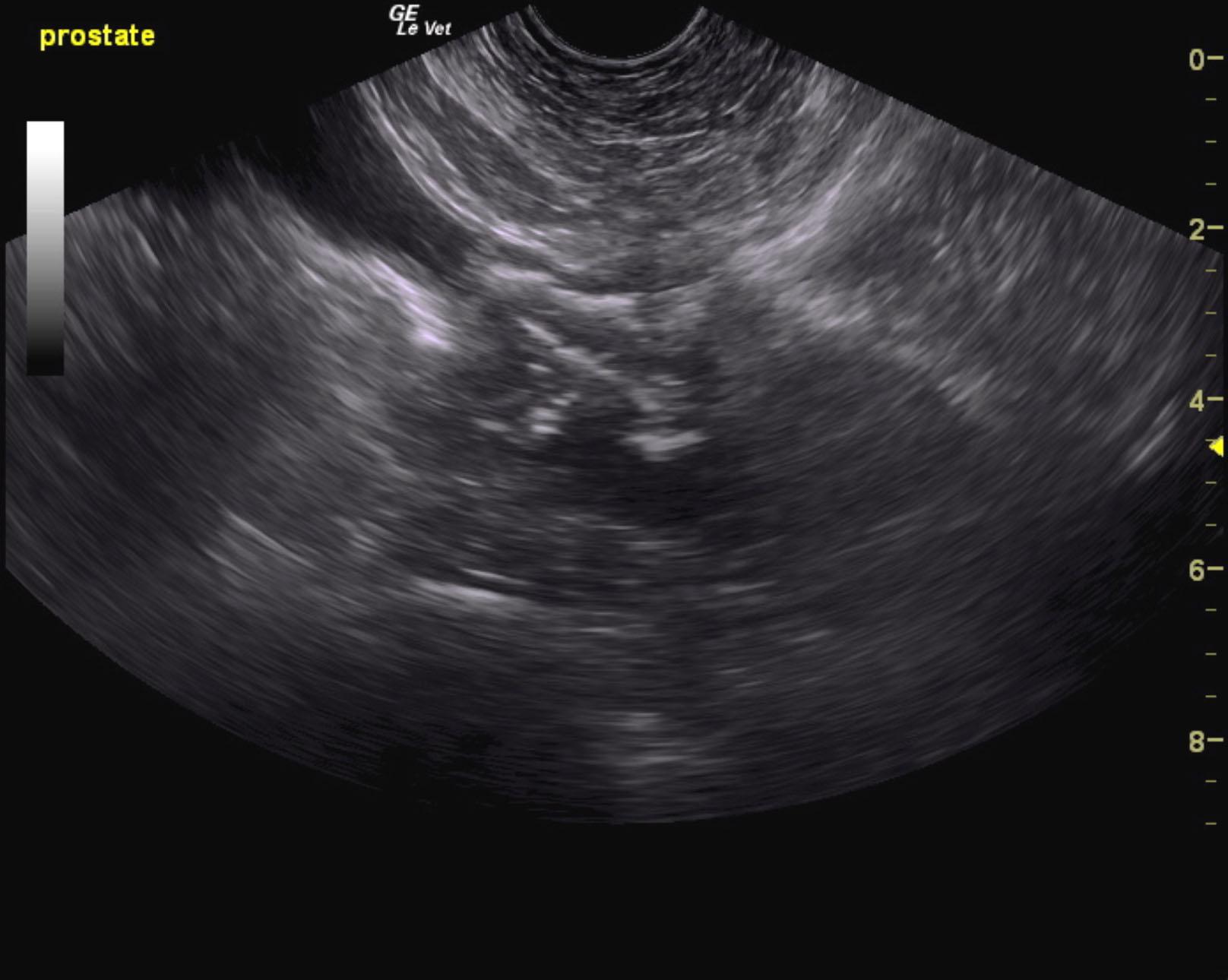
Mineralizing prostatic mass extending into the cystourethral junction.
Iliac trifurcation is free of evident pathology.
No evident metastatic disease. This is strongly suggestive for prostatic carcinoma. Ultrasound-guided FNA are recommended for confirmation. Referral for stent placement at Animal Medical Center with Dr. Weisse and Dr. Berent would be ideal in this case.
The prostate in this patient revealed a mixed, hypoechoic and multifocally mineralizing mass that measured 2.5 cm. Ultrasound-guided FNA or traumatic catheterization is recommended for confirmation. Pericapsular inflammatory pattern was noted around the prostate with proliferative tissue noted cranial to the point of the cystourethral junction.
None
Prostate – neoplasia, abscess, granuloma
None