09_00103 Grady S Bilateral cryptorchid. Unilateral neoplastic testicle

Case Study

09_00103 Grady S Bilateral cryptorchid. Unilateral neoplastic testicle

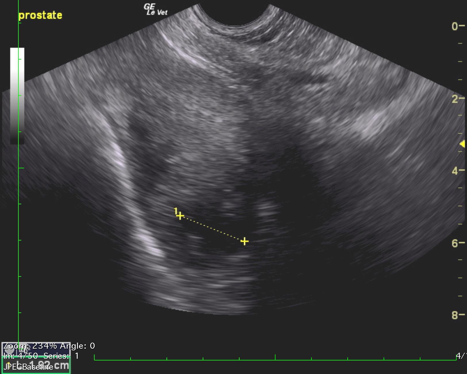
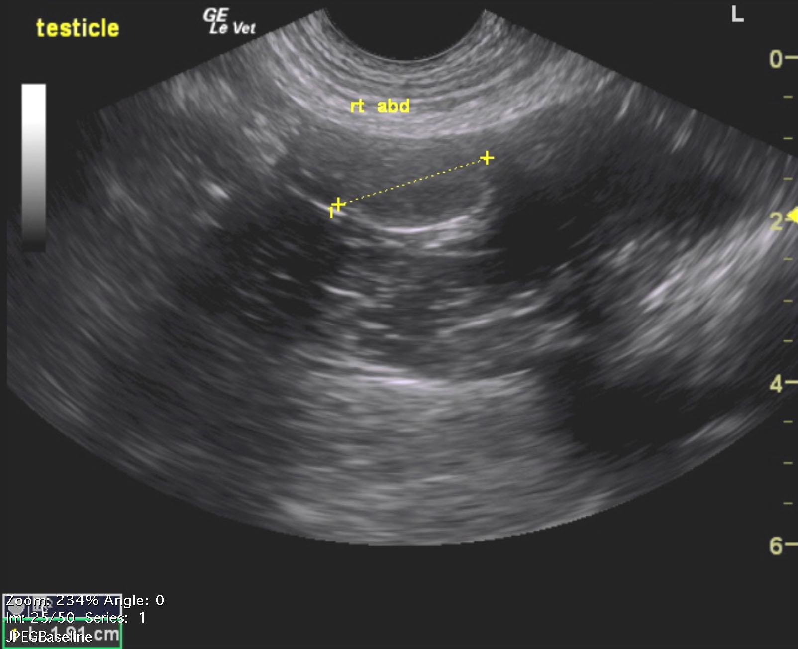
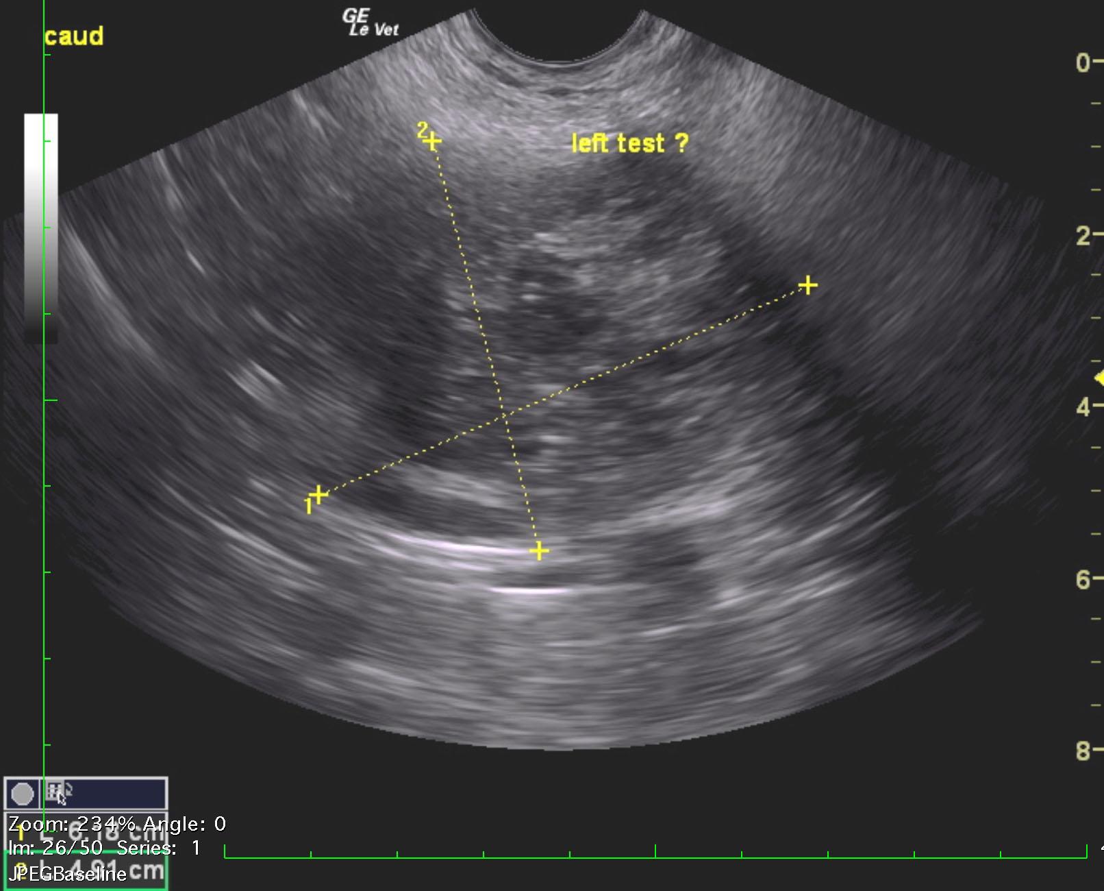
A 9-year-old intact male Boxer was presented for evaluation of blood dripping blood from penis and hematochezia. On physical examination the prepuce area was edematous and reddened, there was a bloody discharge from penis, and a large soft tissue mass was present on rectal palpation. 


Sonographic Differential Diagnosis

Bilateral cryptorchid. Neoplastic transformation of the left testicle.
BPH prostatitis type prostatic pattern with cysts, potential abscessation.

Image Interpretation

The prostate was significantly enlarged, hypoechoic to the surrounding fat. The prostate measured 6.72 x 4.26 cm with cystic changes. Potential abscessation. Periserosal inflammatory pattern was noted. The right testicle was localized within the abdomen, just cranial to the urinary bladder. The right testicle appeared normal in size and contour. The left testicle comprised a mass cranial and left of the urinary bladder and measured 6.18 x 4.9 cm with complex cystic and nodular changes. This is suggestive for Sertoli cell tumor.

DX

Bilateral cryptorchid. Unilateral neoplastic testicle

Outcome

None

Clinical Differential Diagnosis

Prostate – benign hyperplasia, neoplasia, acute prostatitis, chronic prostatitis, abscessation
Colon – neoplasia, granulomatous colitis, abscess

Sampling

None

Patient Information

Gender : Male, Intact
Species : Canine
Type of Imaging : Ultrasound
Status : Complete

Clinical Signs

  • Fresh Blood in Stool

Exam Finding

  • Bleeding from penis
  • Palpable mass

Images

grady_seigel_crypt_sertoli_presumed_prostatomegalygrady_seigel_crypt_right_normalgrady_seigel_crypt_sertoli_presumed

Clinical Signs

  • Fresh Blood in Stool