A 5-year-old NM Pit bull terrier was presented for evaluation of intermittent vomiting. Abnormalities on CBC and serum biochemistry were lymphocytosis (43,900) thrombocytopenia (160) and severe hyperglobulinemia (6.9).
A 5-year-old NM Pit bull terrier was presented for evaluation of intermittent vomiting. Abnormalities on CBC and serum biochemistry were lymphocytosis (43,900) thrombocytopenia (160) and severe hyperglobulinemia (6.9).
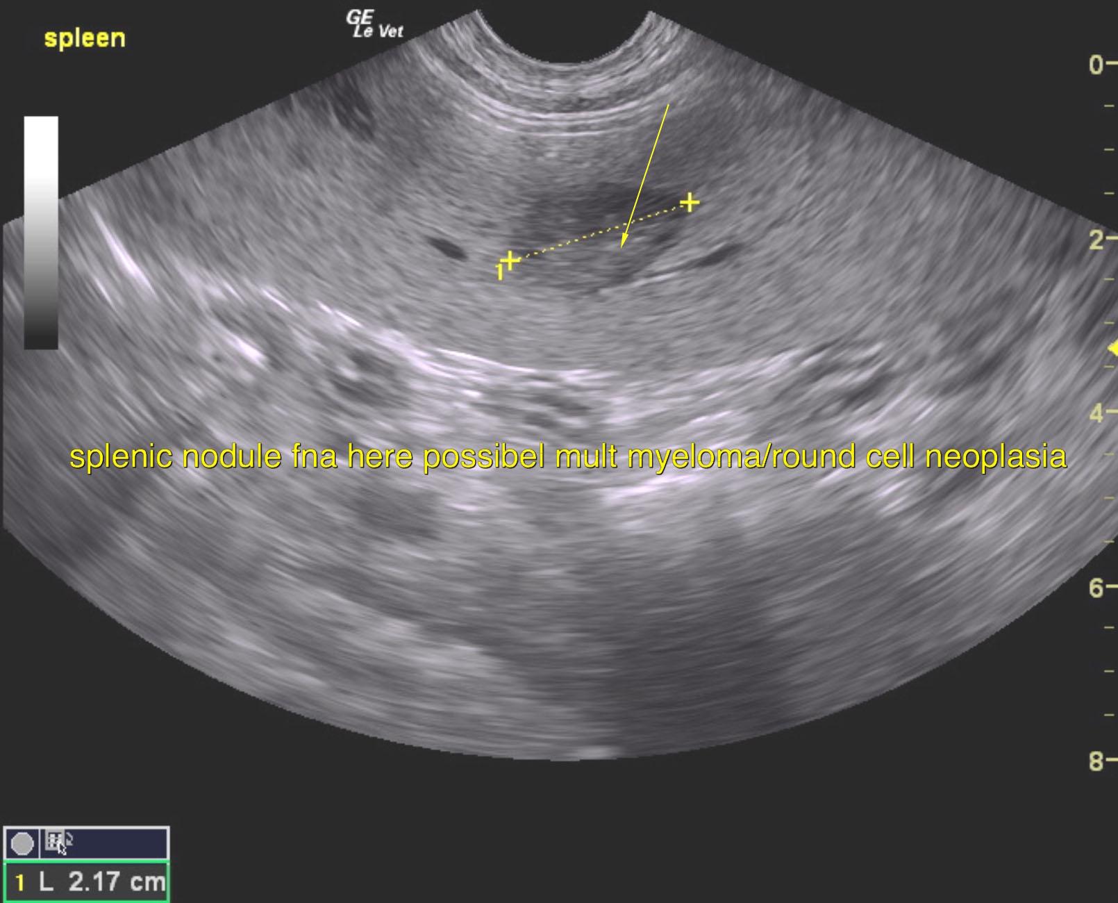
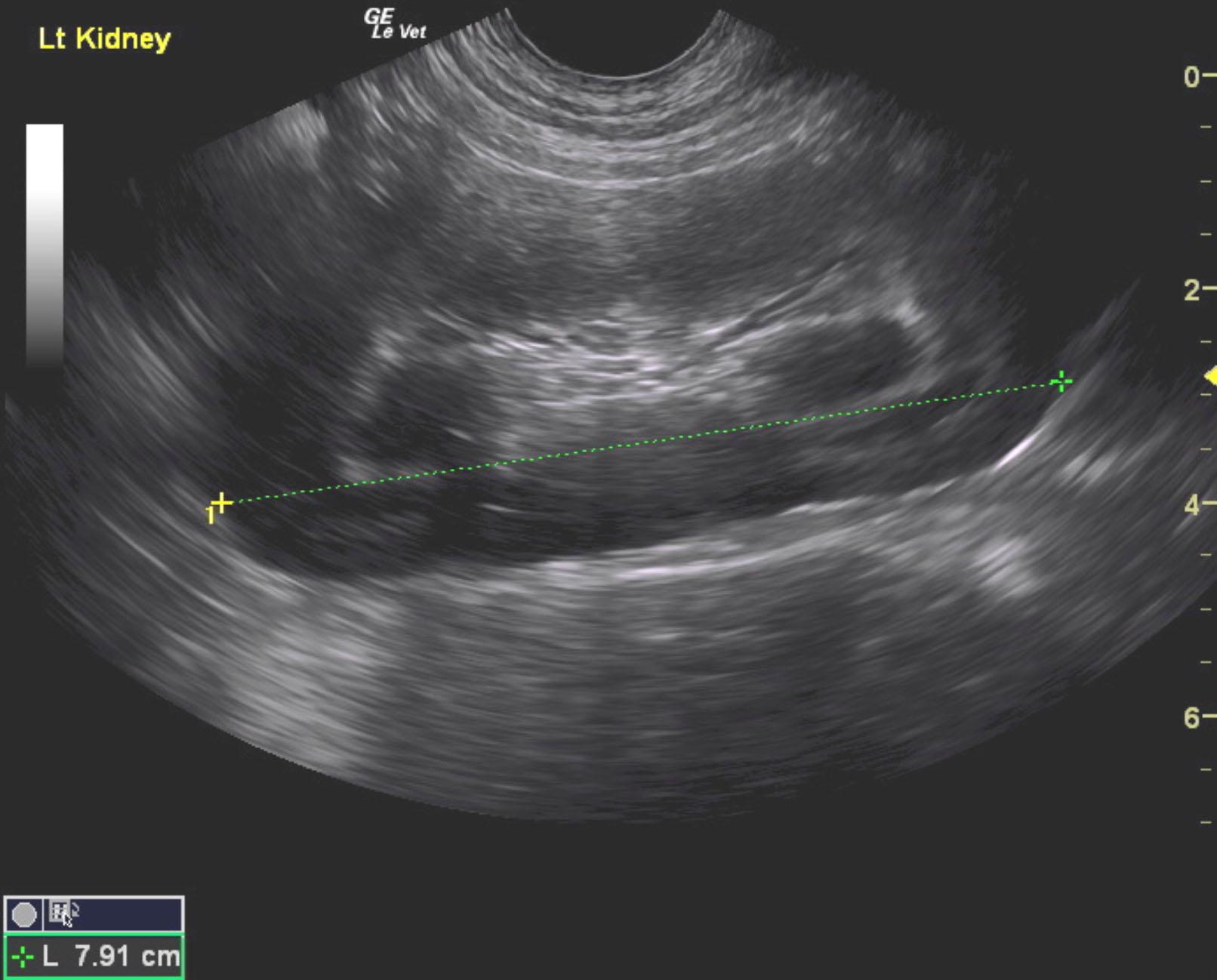
Splenic nodules. Swollen kidneys with medullary rim sign. Suspect round cell neoplasia or multiple myeloma. Ultrasound guided FNA of the spleen would be recommended as well as assessment of the urine for Bence Jones protein given the medullary rim in the kidney and swollen contour. PCR of the blood smear can also be considered for possible lymphoma. Bone marrow biopsy would be ideal in this case. Guarded prognosis.
Spleen revealed focal hypoechoic target-type nodule measuring 2.17 cm. Kidneys were swollen 7.9 cm with medullary rim sign.
None
Multiple myeloma, acute lymphoblastic leukemia, chronic lymphoblastic leukemia, stage V lymphoma, Ehrlichiosis
None