An 11-year-old SF Labrador was presented for evaluation of lethargy and polypnea. On survey radiographs, opacity in the cranial abdomen that displaced the stomach was evident. Urine specific gravity was 1.016. CBC showed leukopenia.
An 11-year-old SF Labrador was presented for evaluation of lethargy and polypnea. On survey radiographs, opacity in the cranial abdomen that displaced the stomach was evident. Urine specific gravity was 1.016. CBC showed leukopenia.
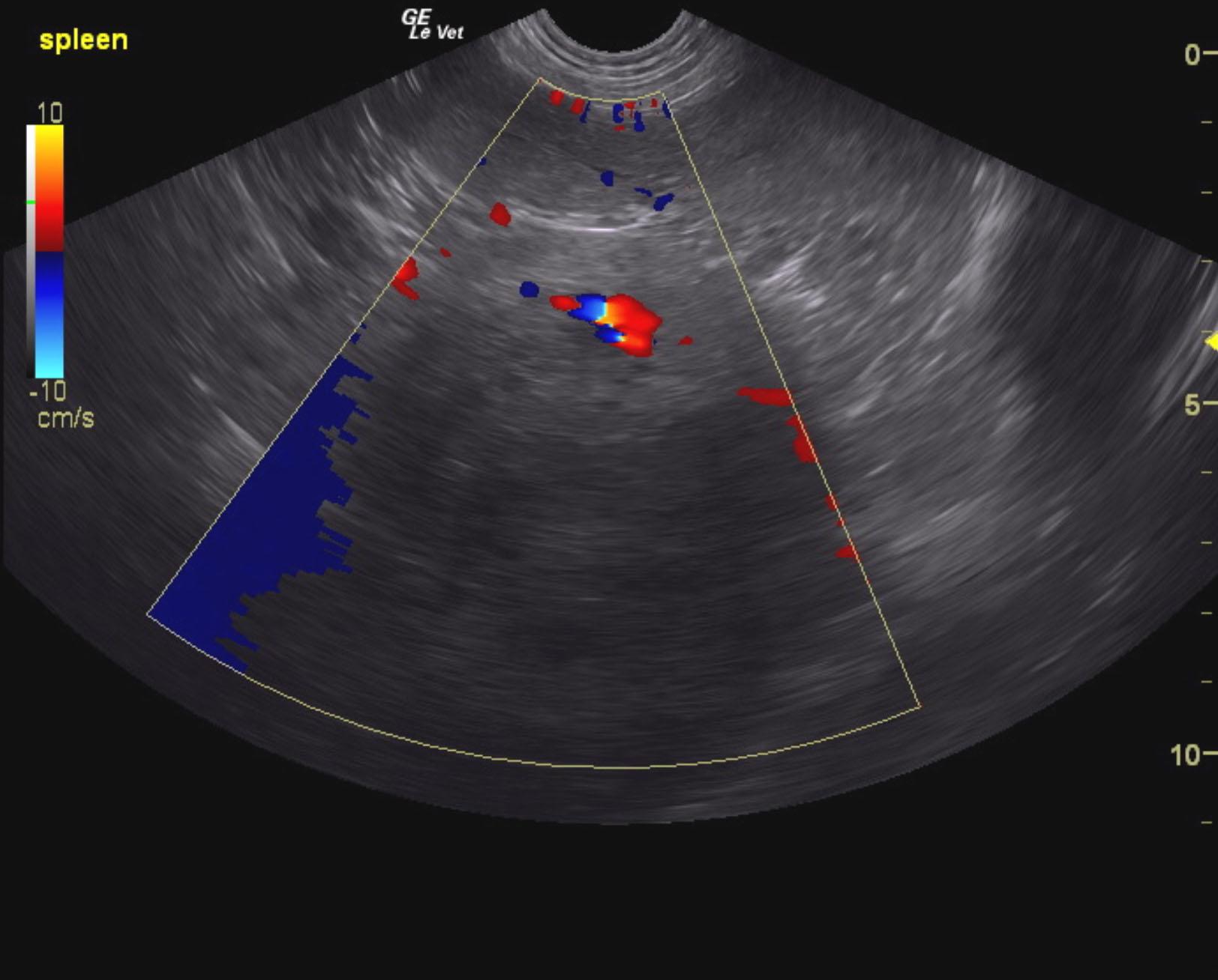
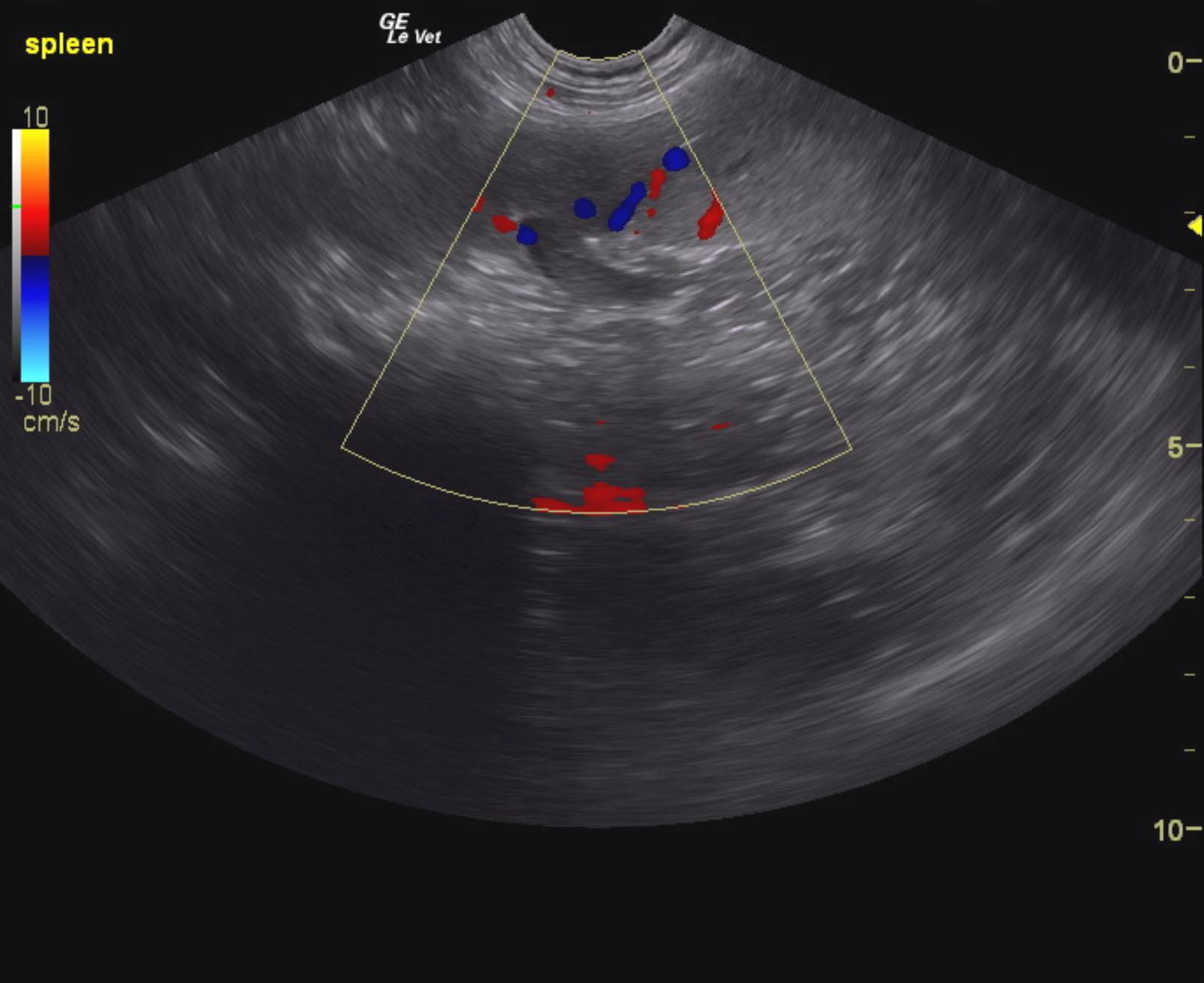
Solitary splenic mass with concurrent splenic thrombus. Splenic hematoma, degenerative process and hemangiosarcoma are all possible.
Chest radiographs and echocardiogram are recommended to assess for metastatic disease.
The spleen presented a moderately complex 9.3 cm mass that was deriving from the mid caudal body. The spleen concurrently demonstrated a thrombus.
The heart revealed no evident pathology.
None
Neoplasia/abscess/granuloma/cyst – spleen, kidney, pancreas, mesentery, lymph node
Splenic torsion
Hydronephrosis
Focal peritonitis
None