08_00151 Yuri G Splenic mass-HSA vs benign

Case Study

08_00151 Yuri G Splenic mass-HSA vs benign

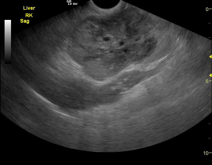
An 11-year-old intact male Yorkshire terrier was presented for evaluation of palpable abdominal mass on routine examination. According to the owner, the dog was clinically normal. Blood work was unremarkable.


Sonographic Differential Diagnosis

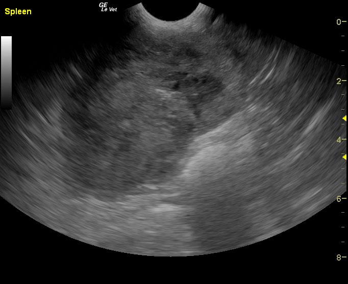
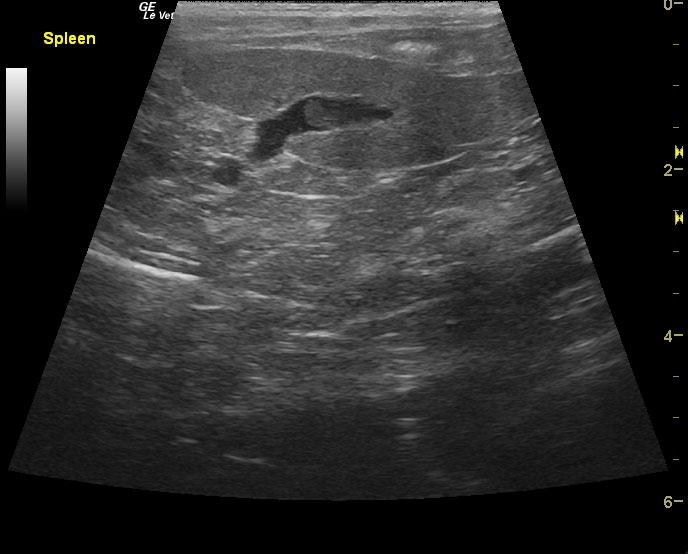
Degenerative hematoma versus hemangiosarcoma or similar neoplasia is suspected with concurrent splenic thrombus.

Image Interpretation

The spleen presented a 6.0 cm, complex, cavitated mass with concurrent splenic thrombosis. This was deriving from the cranial pole.

DX

Splenic mass

Outcome

None

Clinical Differential Diagnosis

Neoplasia/cyst/granuloma/hematoma – liver, spleen, kidney, lymph node, pancreas, mesentery
Hydronephrosis

Sampling

None

Patient Information

Gender : Male, Intact
Species : Canine
Type of Imaging : Ultrasound
Status : Complete

Clinical Signs

  • Palpable Mass

Exam Finding

  • Palpable mass

Images

201507062015352015070620165920150706201119

Clinical Signs

  • Palpable Mass
Skip to content