08_00119 Farley V Splenic thrombus

Case Study

08_00119 Farley V Splenic thrombus

A 13-year-old NM Bassett hound with a history of hepatocellular carcinoma, that was resected in four years ago, was presented for evaluation of weight loss and poor appetite. Abnormalities on serum biochemistry were azotemia and elevated ALT and ALP activity.
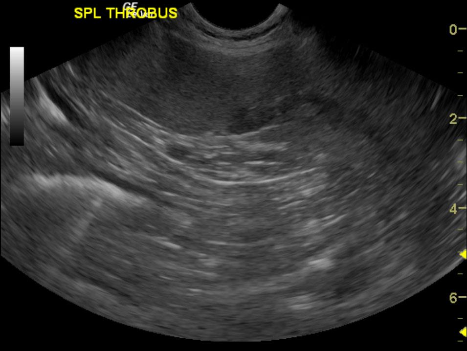
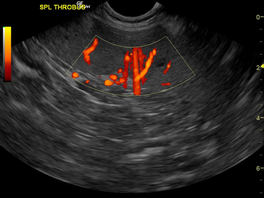
A 13-year-old NM Bassett hound with a history of hepatocellular carcinoma, that was resected in four years ago, was presented for evaluation of weight loss and poor appetite. Abnormalities on serum biochemistry were azotemia and elevated ALT and ALP activity.

Sonographic Differential Diagnosis

Splenic thrombus. No overt evidence of neoplasia.
Assessment for infectious disease and causes of vasculitis should be investigated. Full coagulation panel, Clindamycin or Doxycycline, Plavix and or aspirin at 1 mg/kg s.i.d. would be recommended. A recheck sonogram in 1 week primarily of the spleen is recommended.

Image Interpretation

The spleen was largely uniform. However, a splenic thrombus was noted at the caudal branch of the spleen entering into the main splenic vein at approximately 4.0 cm. This area of the splenic vein was void of power Doppler signal.

DX

Splenic thrombus

Outcome

None

Clinical Differential Diagnosis

Liver – neoplasia, nodular regeneration, vacuolar hepatopathy, infectious (bacterial, fungal, viral), toxins
Renal – chronic kidney disease, neoplasia, pyelonephritis, renoliths

Sampling

None

Patient Information

Gender : Male, Neutered
Species : Canine
Type of Imaging : Ultrasound

Clinical Signs

  • Anorexia
  • Weight loss

Images

spleen_farley_vitalo_k9basset13yomnak_splenic_thrombus_2spl_thrombus_doppler

Blood Chemistry

  • Alkaline Phosphatase (SAP), High
  • ALT (SGPT), High
  • Azotemia

Clinical Signs

  • Anorexia
  • Weight loss