Skip to content
Customize Consent Preferences

We use cookies to help you navigate efficiently and perform certain functions. You will find detailed information about all cookies under each consent category below.

The cookies that are categorized as "Necessary" are stored on your browser as they are essential for enabling the basic functionalities of the site. ... 

Always Active

Necessary cookies are required to enable the basic features of this site, such as providing secure log-in or adjusting your consent preferences. These cookies do not store any personally identifiable data.

No cookies to display.

Functional cookies help perform certain functionalities like sharing the content of the website on social media platforms, collecting feedback, and other third-party features.

No cookies to display.

Analytical cookies are used to understand how visitors interact with the website. These cookies help provide information on metrics such as the number of visitors, bounce rate, traffic source, etc.

No cookies to display.

Performance cookies are used to understand and analyze the key performance indexes of the website which helps in delivering a better user experience for the visitors.

No cookies to display.

Advertisement cookies are used to provide visitors with customized advertisements based on the pages you visited previously and to analyze the effectiveness of the ad campaigns.

No cookies to display.

08-00137 Dina N Splenic mass—NEED SL——-

Case Study

08-00137 Dina N Splenic mass—NEED SL——-

A 7-year-old SF Labradoodle was presented for evaluation of lethargy, abdominal distension, and elevated respiratory and heart rate. Abnormalities on CBC and serum biochemistry were mild leukocytosis, hypoalbuminemia, and elevated ALP activity (181).


Sonographic Differential Diagnosis

Splenic mass. FNAs of the splenic mass were performed without complication.
Deep left medial liver nodule. Hyperplasia versus metastatic lesion.
FNAs of the liver nodule were performed without complication.

Image Interpretation

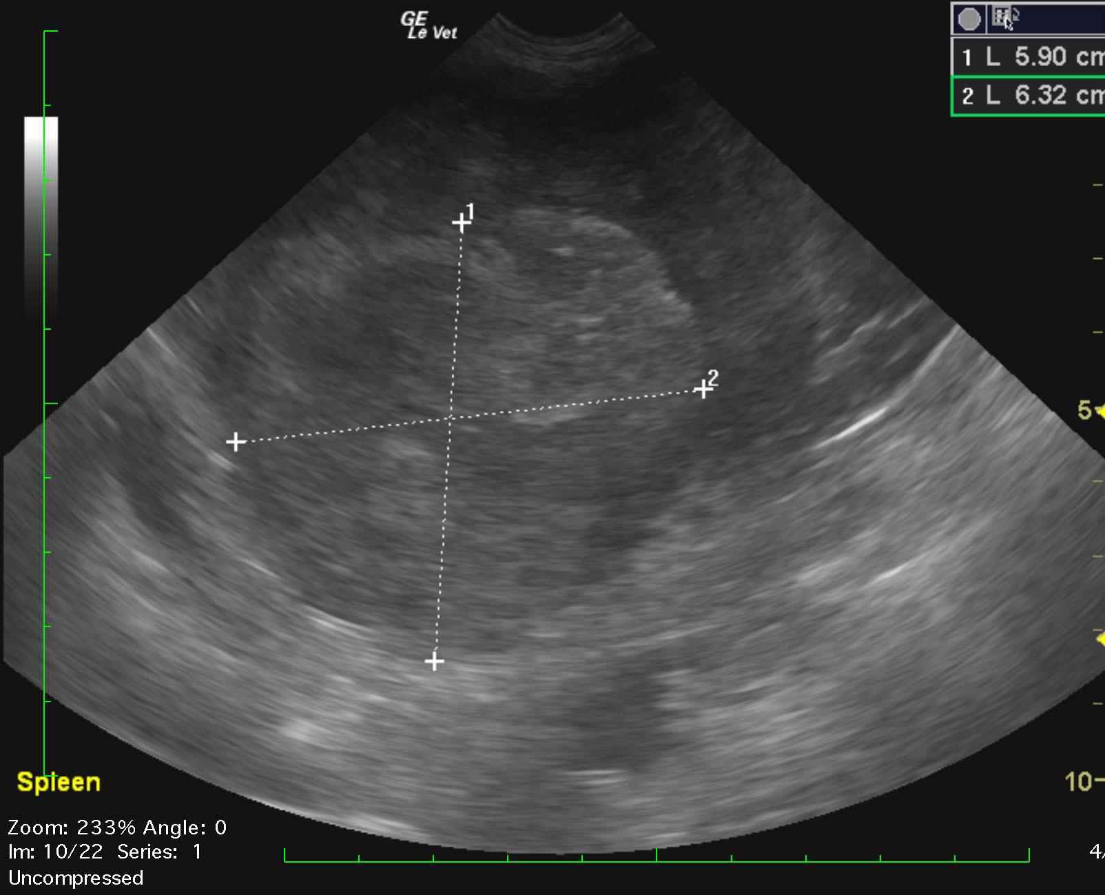
The spleen revealed a mixed, hyperechoic mass that measured 6.3 x 5.9 cm at the cranial pole. Coalesced free fluid was noted associated with the splenic mass. Minor reactive surrounding fat was noted. A hyperechoic, right medial liver nodule measured 1.24 x 1.17 cm. This is most consistent with lipogranuloma. Left medial liver nodule was mildly echogenic and measured 1.66 x 1.61 cm. Neither nodule revealed disruption of architecture. The left medial liver nodule could represent a metastatic event. Mild, uniform hepatic swelling was noted. No significant hepatic lymphadenopathy was noted.

Outcome

None

Clinical Differential Diagnosis

Cardiac – cardiomyopathy, pericardial effusion
Neoplasia – spleen, liver
Peritonitis

Sampling

??

Video

Patient Information

Gender : Female, Spayed
Species : Canine
Type of Imaging : Ultrasound

Clinical Signs

  • Abdominal Distension
  • Lethargy
  • Tachypnea

Exam Finding

  • Abdominal Distension
  • Tachycardia
  • Tachypnea

Images

dina_nichols_splenic_mass_liver_nodule_fnadina_nichols_splenic_mass_liver_nodule_1

Blood Chemistry

  • Albumin, Low

CBC

  • WBC, High

Clinical Signs

  • Abdominal Distension
  • Lethargy
  • Tachypnea