Skip to content
Customize Consent Preferences

We use cookies to help you navigate efficiently and perform certain functions. You will find detailed information about all cookies under each consent category below.

The cookies that are categorized as "Necessary" are stored on your browser as they are essential for enabling the basic functionalities of the site. ... 

Always Active

Necessary cookies are required to enable the basic features of this site, such as providing secure log-in or adjusting your consent preferences. These cookies do not store any personally identifiable data.

No cookies to display.

Functional cookies help perform certain functionalities like sharing the content of the website on social media platforms, collecting feedback, and other third-party features.

No cookies to display.

Analytical cookies are used to understand how visitors interact with the website. These cookies help provide information on metrics such as the number of visitors, bounce rate, traffic source, etc.

No cookies to display.

Performance cookies are used to understand and analyze the key performance indexes of the website which helps in delivering a better user experience for the visitors.

No cookies to display.

Advertisement cookies are used to provide visitors with customized advertisements based on the pages you visited previously and to analyze the effectiveness of the ad campaigns.

No cookies to display.

07_00181 CD C Unilateral adrenal mass, +LDDS, nl contralateral adrenal

Case Study

07_00181 CD C Unilateral adrenal mass, +LDDS, nl contralateral adrenal

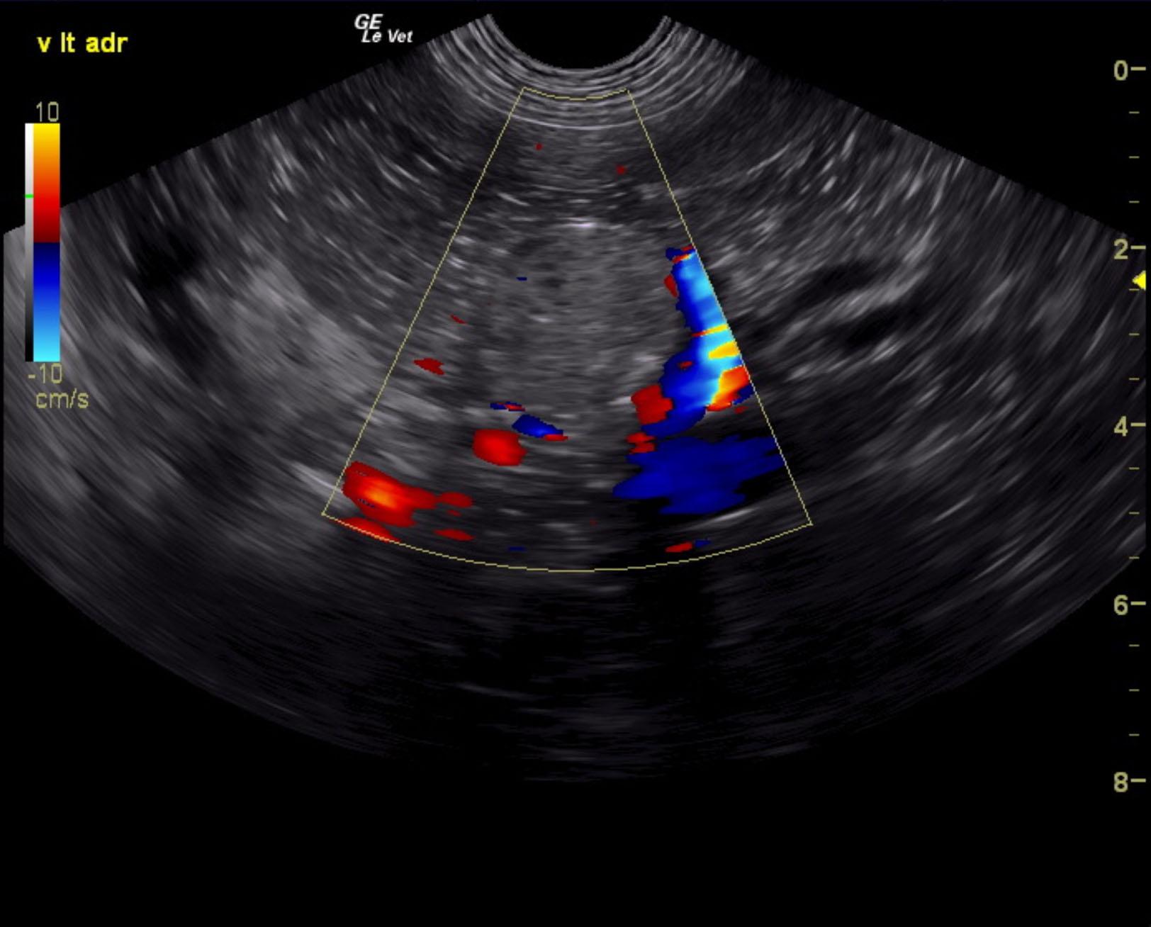
A 7-year-old SF mixed breed dog was presented for evaluation of PU/PD and polypnea. On physical examination the fur on the flanks was bilaterally thin and she had a pot-bellied appearance. Urinalysis showed SG of 1.023 and 2+ protein. Abnormalities on serum biochemistry were mildly elevated ALT activity (150 U/l). Low-dose dexamethasone suppression showed cortisol of: pre 5.8, 4 hour 5.4; and 8 hour 4.8 mg/dl.


Sonographic Differential Diagnosis

Left adrenal mass, focal mineralization suggestive for carcinoma. Given the LDDST results functional adenocarcinoma is suspected.

Image Interpretation

Right adrenal uniform 2.55 x 0.62 cm cranial; 0.55 cm caudal. Left adrenal comprised a mass measuring 4 x 2.6 cm caudal; 0.72 cm cranial. Capsular expansion noted without capsular escape. No overt vascular invasion noted. This appears to be resectable. The caudal mass portion of the adrenal measured 2.6 x 0.53 cm deviating the left renal artery caudally.

DX

Functional adrenal mass

Outcome

None

Clinical Differential Diagnosis

Pituitary-dependent Cushing’s disease, adrenal adenoma, adrenal carcinoma

Sampling

None

UA Specific Gravity Range

1.023

Patient Information

Gender : Female, Spayed
Species : Canine
Type of Imaging : Ultrasound

Clinical Signs

  • PU-PD
  • Tachypnea

Exam Finding

  • Alopecia
  • Pot belly

Images

cd2cd3cd4cd5

Blood Chemistry

  • ALT (SGPT), High

Clinical Signs

  • PU-PD
  • Tachypnea

Special Testing

  • LDDST Positive for Cushing's

Urinalysi

  • Protein Present