An 11-year-old SF Shih Tzu was presented for evaluation of a single episode of vomiting and anorexia for 3-4 days. Abnormalities on CBC and serum biochemistry were hemoconcentration, elevated ALT (136) and ALP (1197) activity. Urine SG was 1.035.
An 11-year-old SF Shih Tzu was presented for evaluation of a single episode of vomiting and anorexia for 3-4 days. Abnormalities on CBC and serum biochemistry were hemoconcentration, elevated ALT (136) and ALP (1197) activity. Urine SG was 1.035.
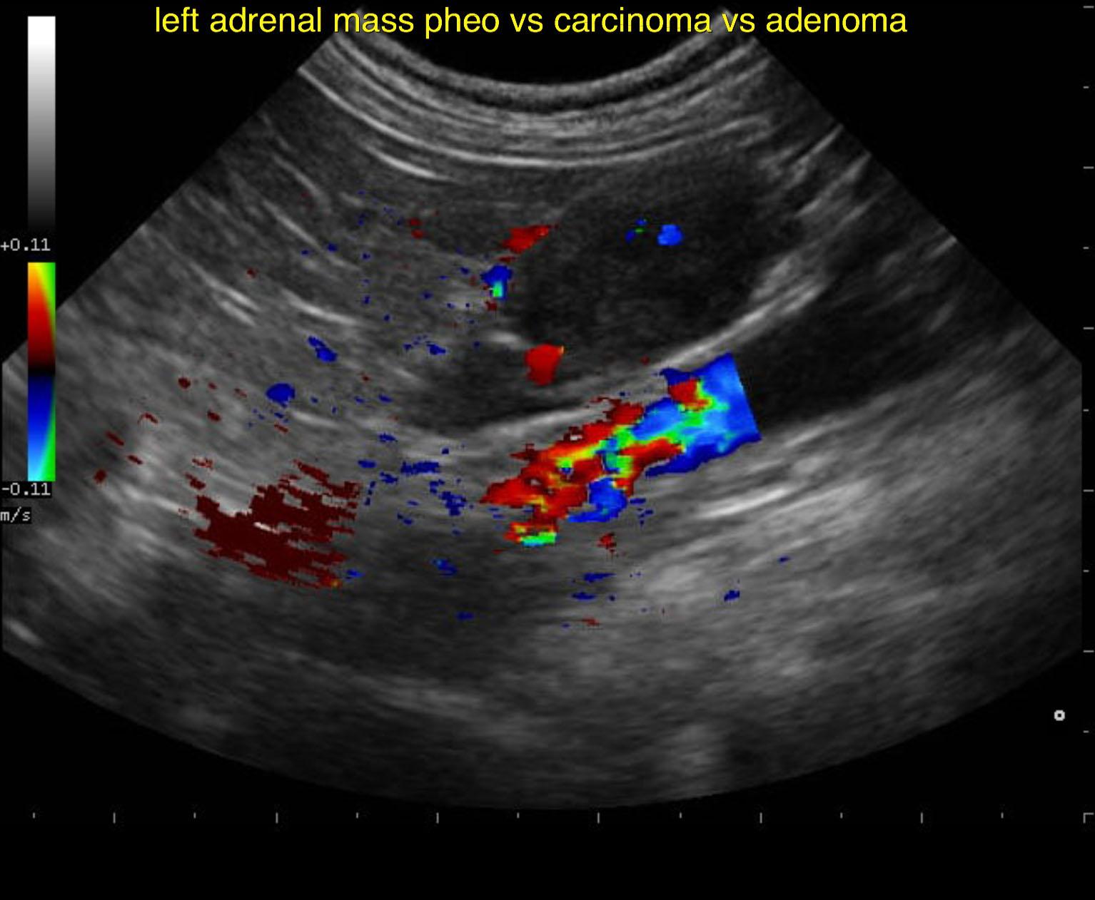
Left adrenal gland mass, non invasive, appears resectable. Differentials include adenoma, pheochromocytoma or adenocarcinoma, which is likely non functional given that isosthenuria is not present. The left adrenal mass appeared moderately vascular. Therefore, pheochromocytoma or adenocarcinoma are significant potentials.
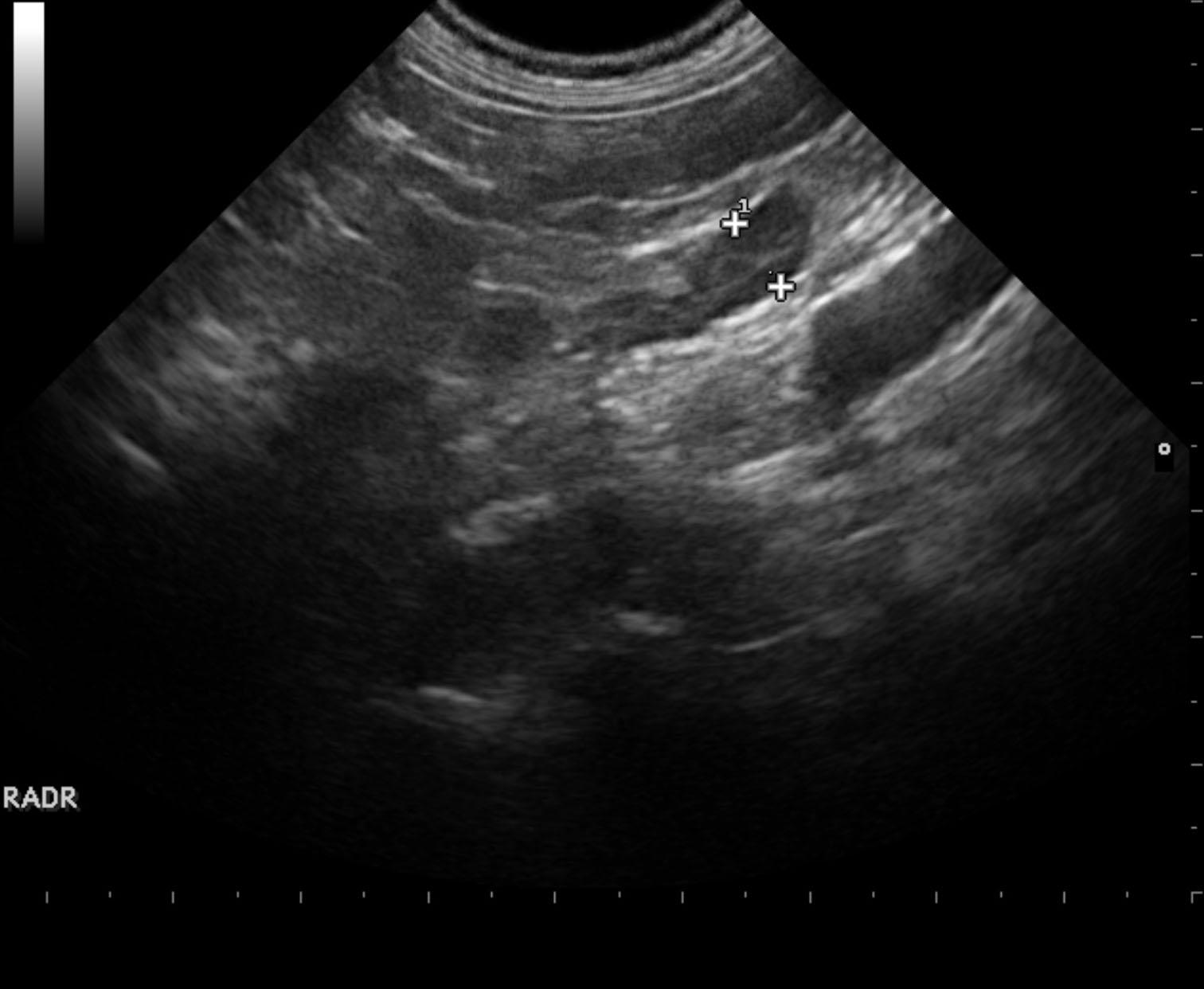
The left adrenal gland was enlarged, nodular and irregular measuring 1.42 cm. The left adrenal gland mass derives from the caudal pole. The left adrenal mass appeared moderately vascular. The right adrenal gland was uniform and measured 0.61 cm in width.
None
Liver – acute hepatitis (viral, bacterial, fungal, toxins), neoplasia, chronic-active hepatitis, cirrhosis
Gall bladder – mucocele, cholecystitis
Adrenal – neoplasia, hyperplasia
None