A 12-year-old NM DSH was presented for evaluation of a palpable abdominal mass that was progressively getting bigger. Urine specific gravity was 1.016 and abnormalities on CBC and serum biochemistry were anemia and elevated fPL.
A 12-year-old NM DSH was presented for evaluation of a palpable abdominal mass that was progressively getting bigger. Urine specific gravity was 1.016 and abnormalities on CBC and serum biochemistry were anemia and elevated fPL.
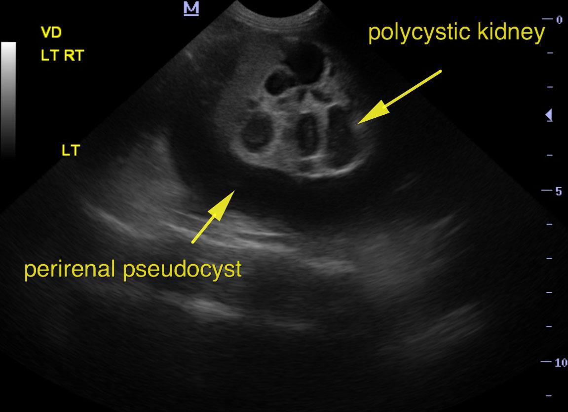
Perirenal pseudocysts, severe on the right and mild to moderate on the left.
Concurrent polycystic renal disease.
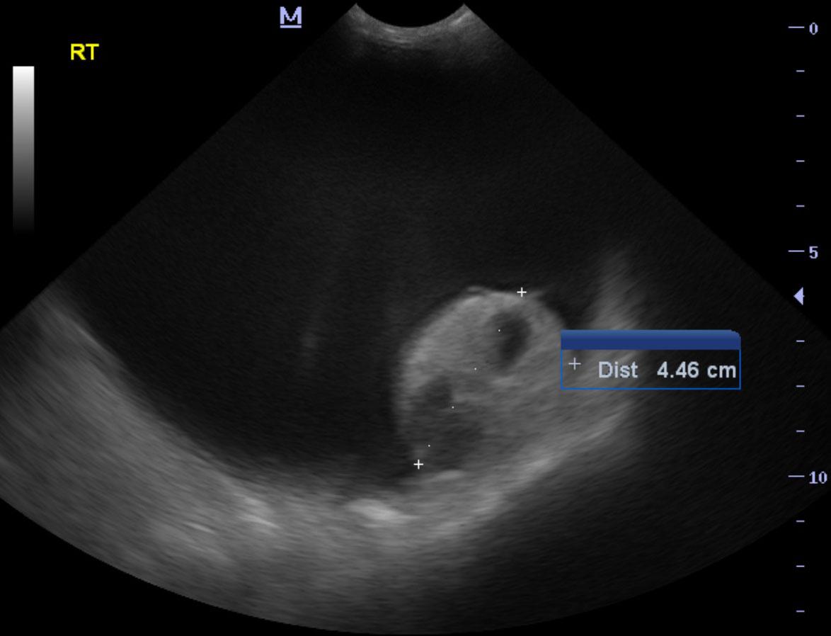
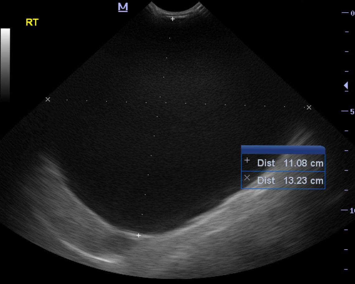
Perirenal pseudocysts and polycystic kidney changes were noted in both kidneys. Disruption of architecture was noted throughout the kidney. The right kidney in this patient presented a large, perirenal pseudocyst with a 4.46 cm dystrophic, interstitial nephrosis pattern. The right kidney including the perirenal pseudocyst extended for approximately 13.0 cm. The left kidney measured 4.63 cm.
None
Mass – neoplasia/cyst/abscess/granuloma of liver, spleen, pancreas, kidney, lymph node, mesentery, abdominal wall
Hydronephrosis
None