A 15-year-old NM DSH with a history of hyperthyroidism, grade 2 heart murmur, and chronic upper respiratory tract disease was presented for evaluation of a palpable abdominal mass. Azotemia was present on serum biochemistry.
A 15-year-old NM DSH with a history of hyperthyroidism, grade 2 heart murmur, and chronic upper respiratory tract disease was presented for evaluation of a palpable abdominal mass. Azotemia was present on serum biochemistry.
Right perirenal pseudocyst.
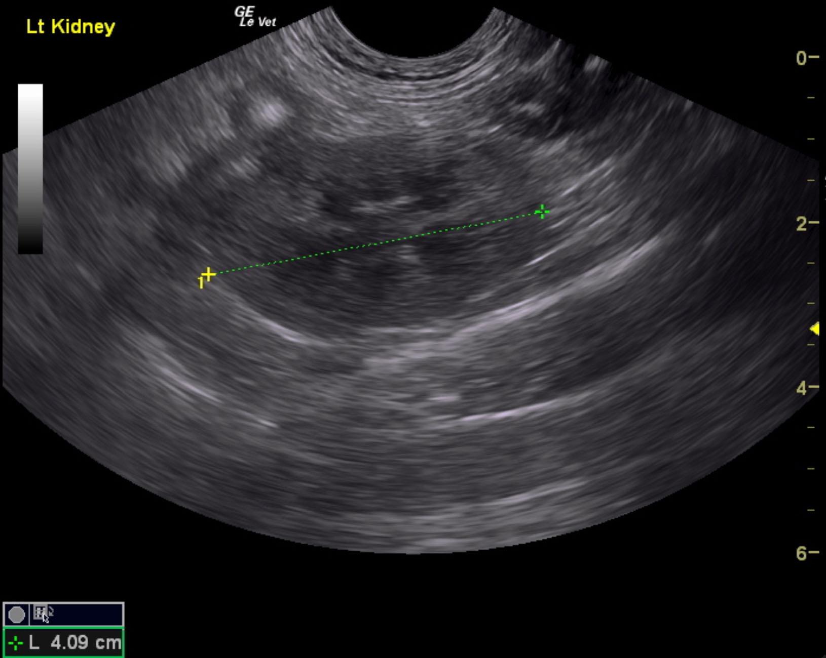
Mildly degenerative left kidney changes.
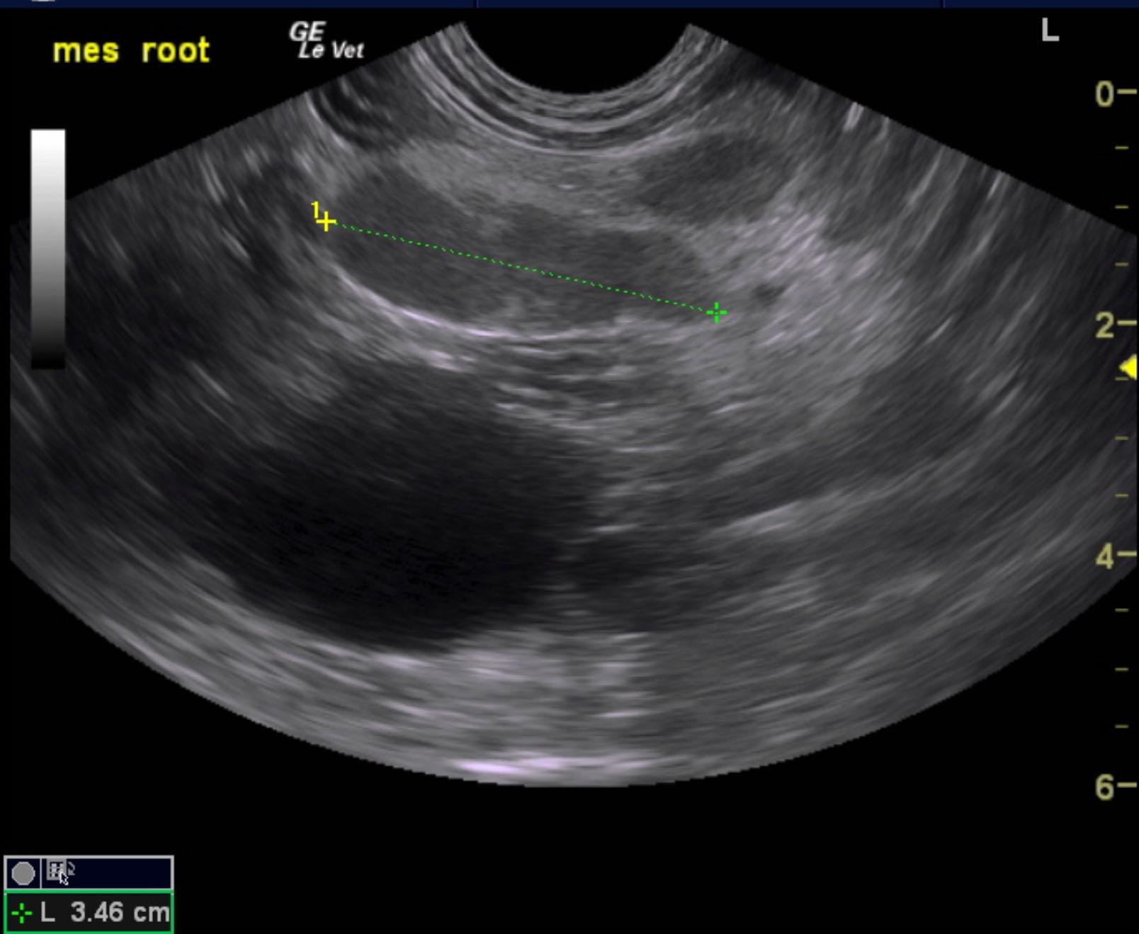
Concurrent mesenteric lymphadenopathy.
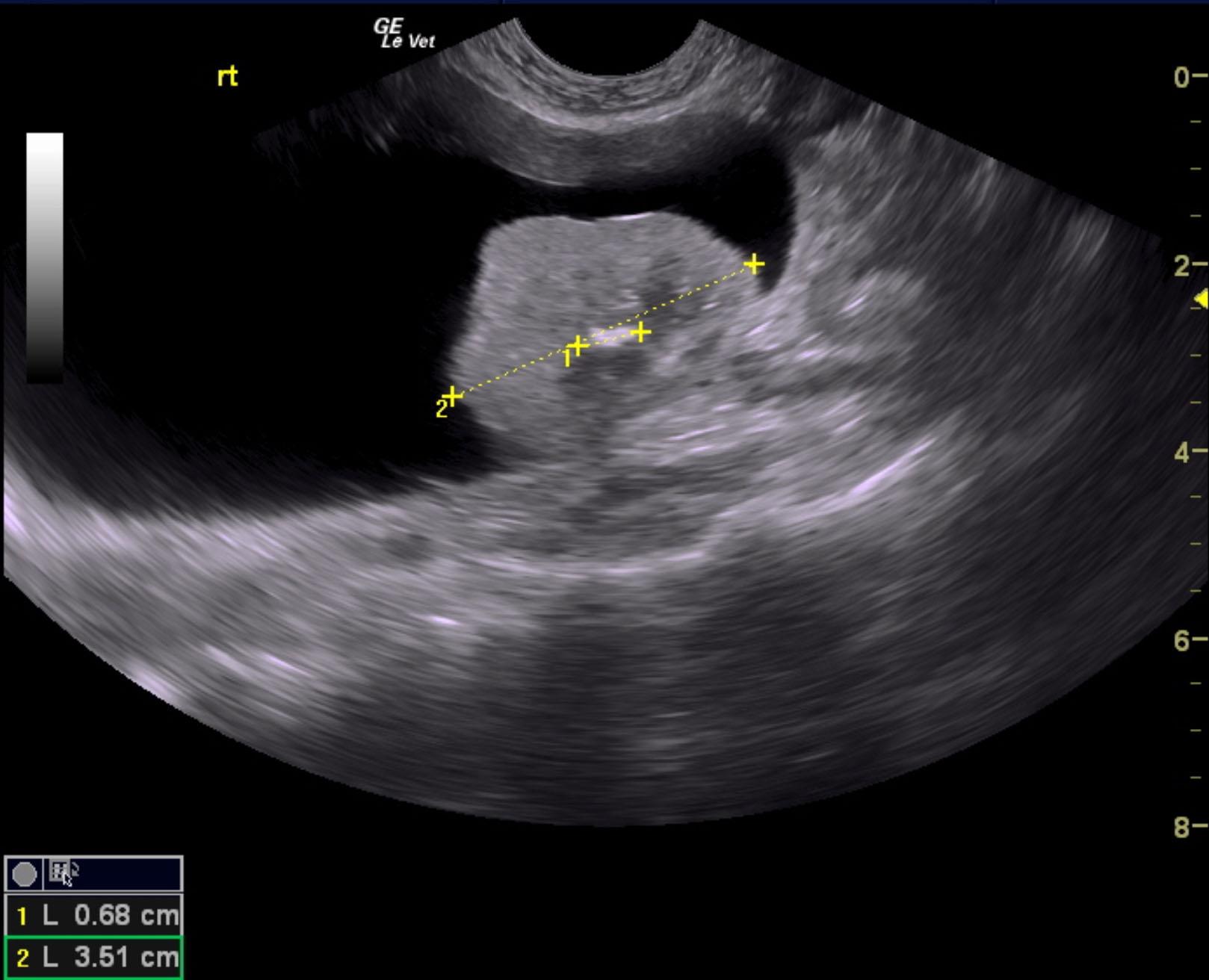
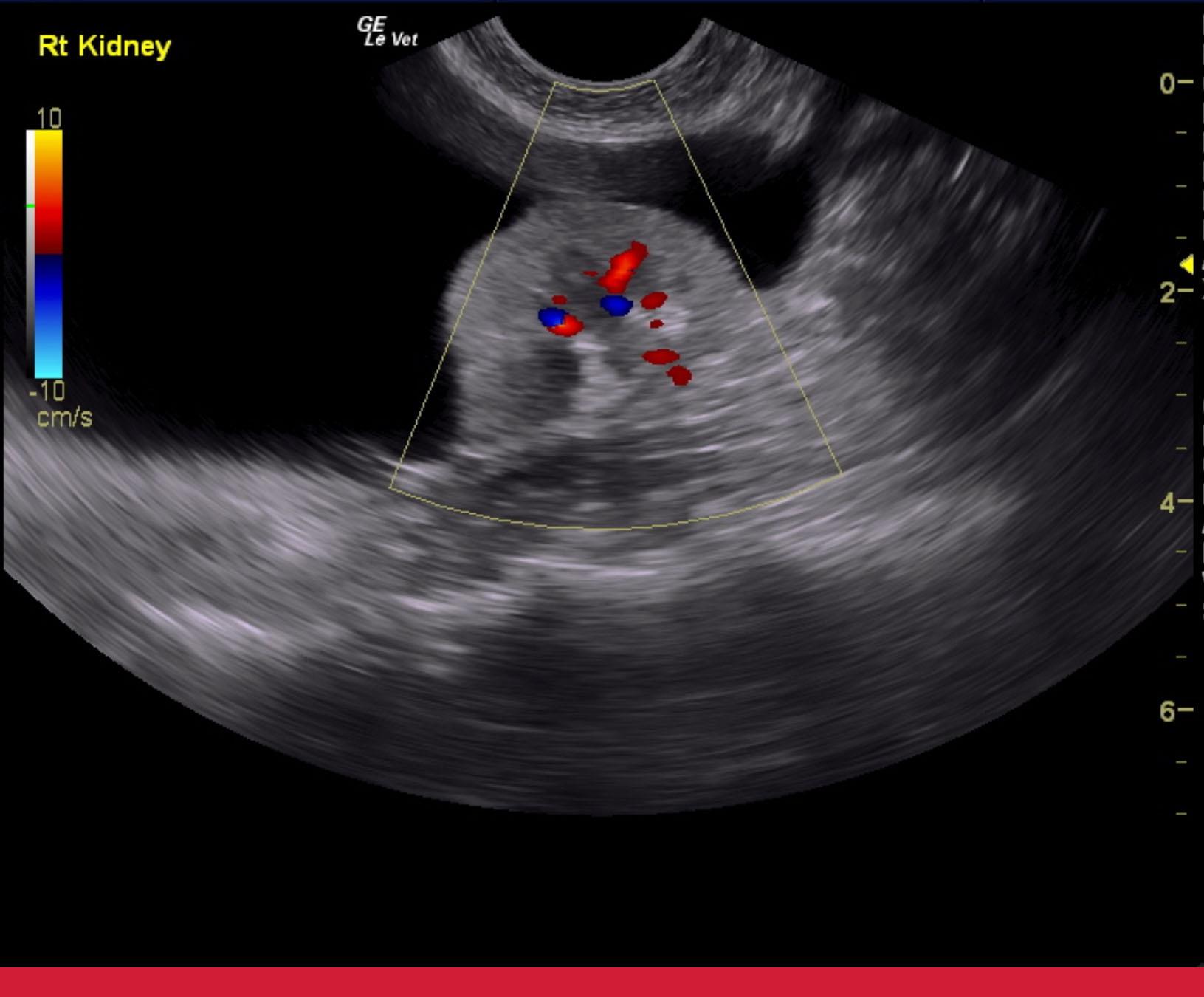
The right kidney in this patient presented chronic interstitial nephrosis pattern with irregular contour and focal, cortical collapse. Pelvic calculus was noted that measured 0.68 cm. The parenchymal portion of the right kidney measured 3.58 cm, yet a perirenal pseudocyst was noted with mild, echogenic debris. The pseudocyst extended for approximately 8.0 cm. Slight pyelectasia of the right kidney was noted and measured 0.23 cm. Color flow of the right kidney appeared to be subjectively subnormal. The left kidney was largely unremarkable with mild degenerative changes with a slightly thickened cortex and measured 4.09 cm. Mesenteric lymph nodes were enlarged and measured 3.4 x 1.0 cm. Irregular contour was noted. FNA is recommended
None
Renal – neoplasia, hydronephrosis, renoliths, pyelonephritis, renal cyst
Mass – neoplasia/cyst/abscess/granuloma of liver, spleen, pancreas, lymph node, mesentery, abdominal wall
None