A 3-year-old NM Goldendoodle was presented for evaluation for hematuria. Survey radiographs showed a mid-abdominal mass effect.
A 3-year-old NM Goldendoodle was presented for evaluation for hematuria. Survey radiographs showed a mid-abdominal mass effect.
Left renal mass appears isolated.
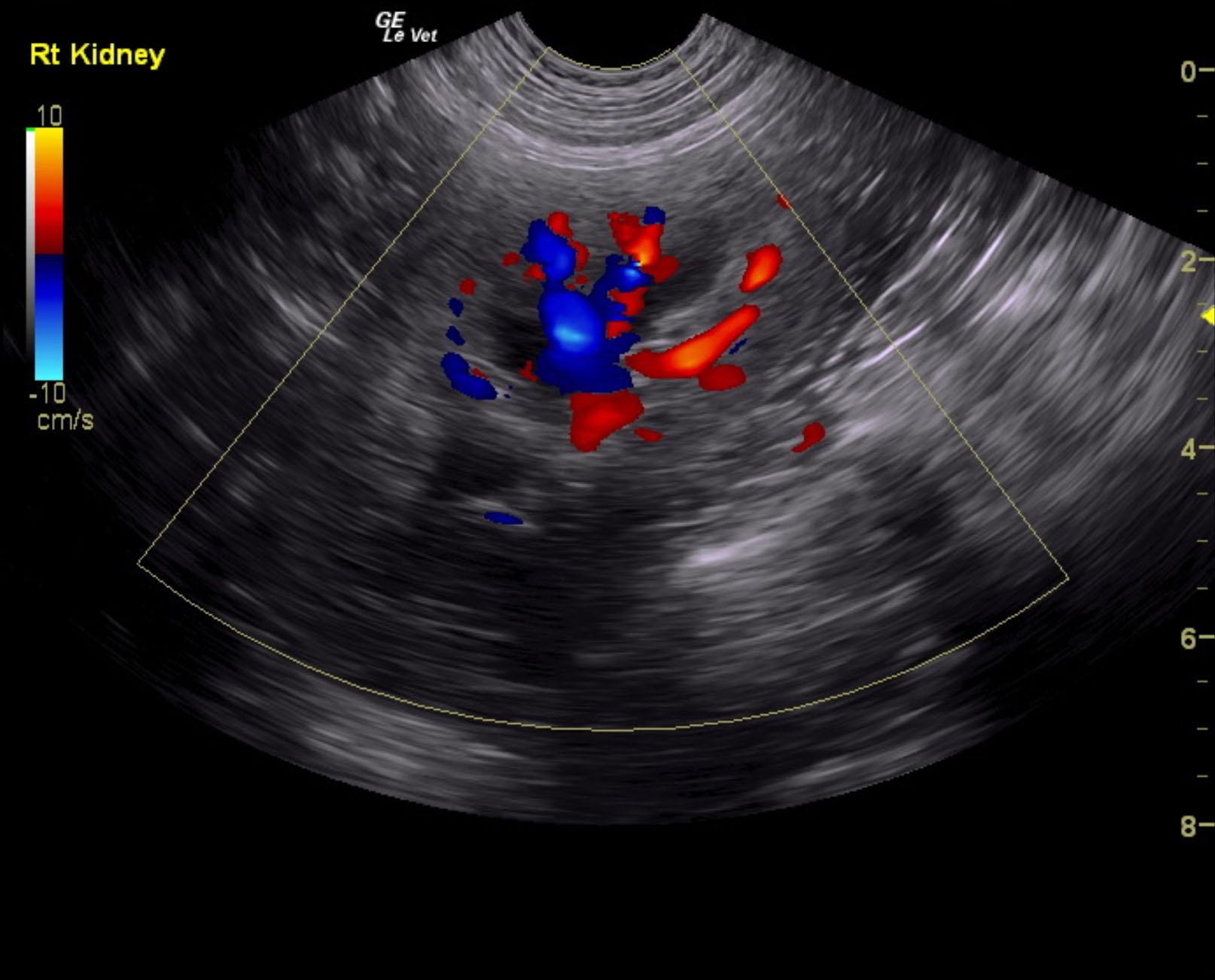
Unremarkable right kidney, suspect renal carcinoma, possible round cell neoplasia.
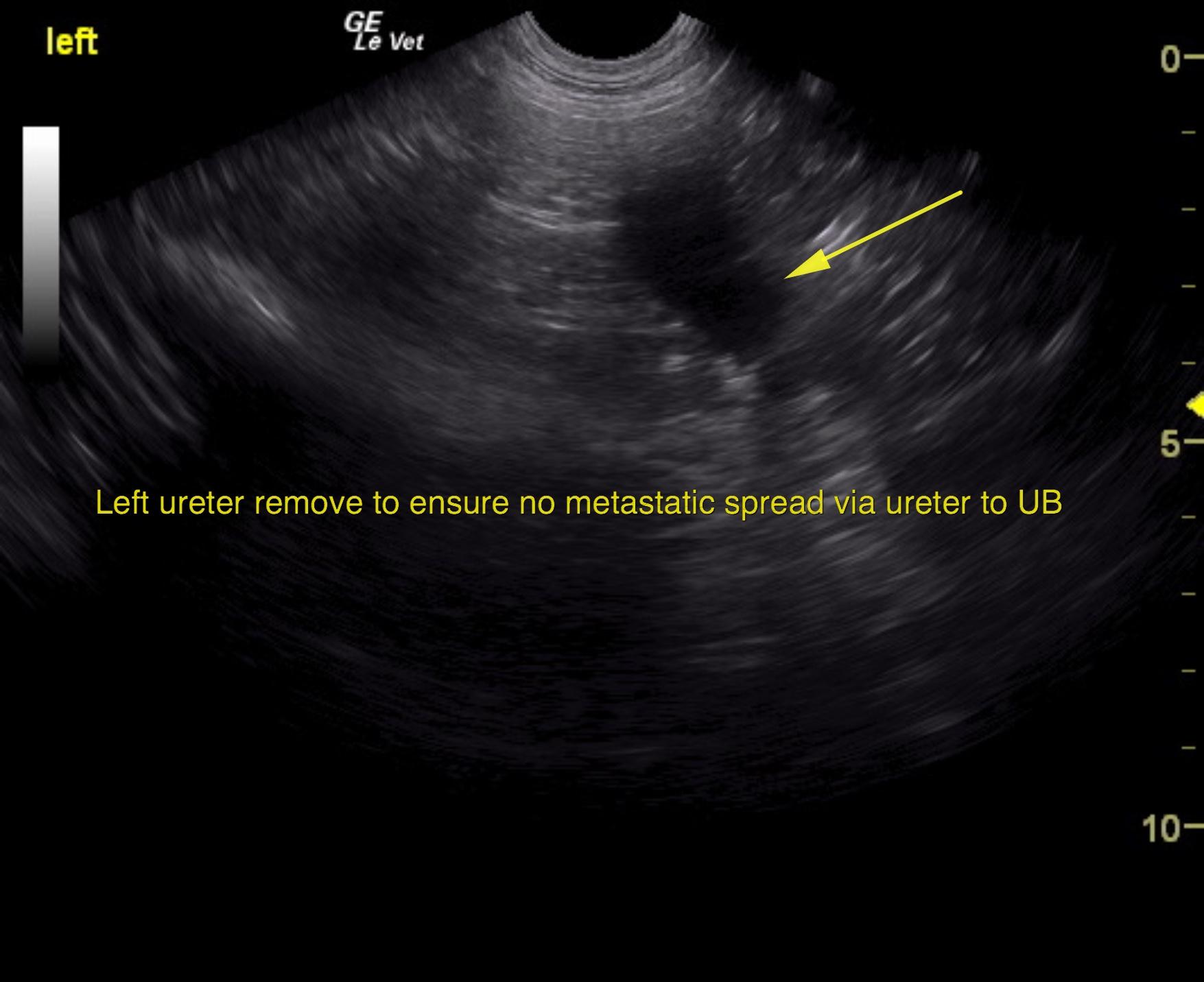
Given that the mass appears to be encapsulated I would recommend utilizing the renal capsule to the benefit of this patient by performing direct left nephrectomy as long as chest radiographs are free of evident pathology and no evidence of CNS disease is present. Immediate left nephrectomy is recommended. Left ureterectomy is recommended to ensure that metastasis through the ureter is not an issue. The ureter should be removed to the level of the urinary bladder.
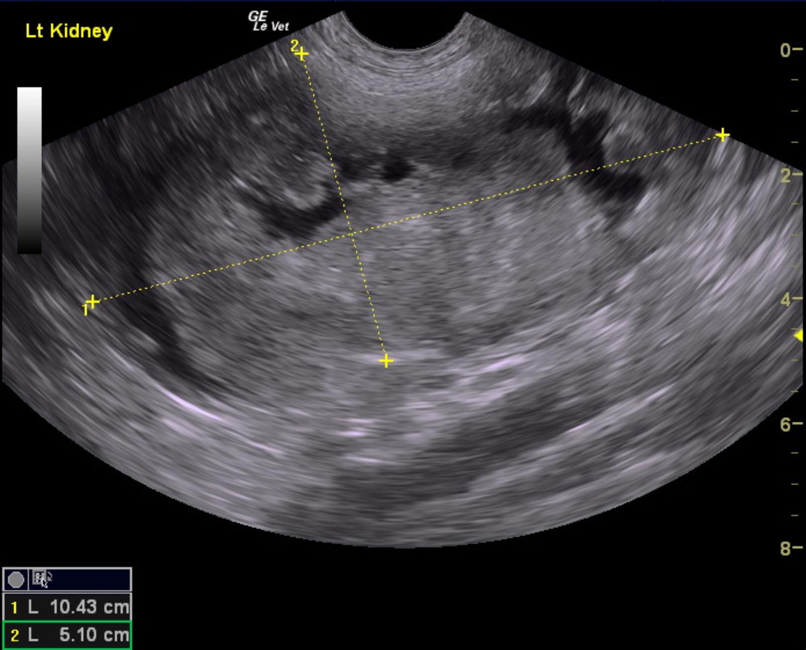
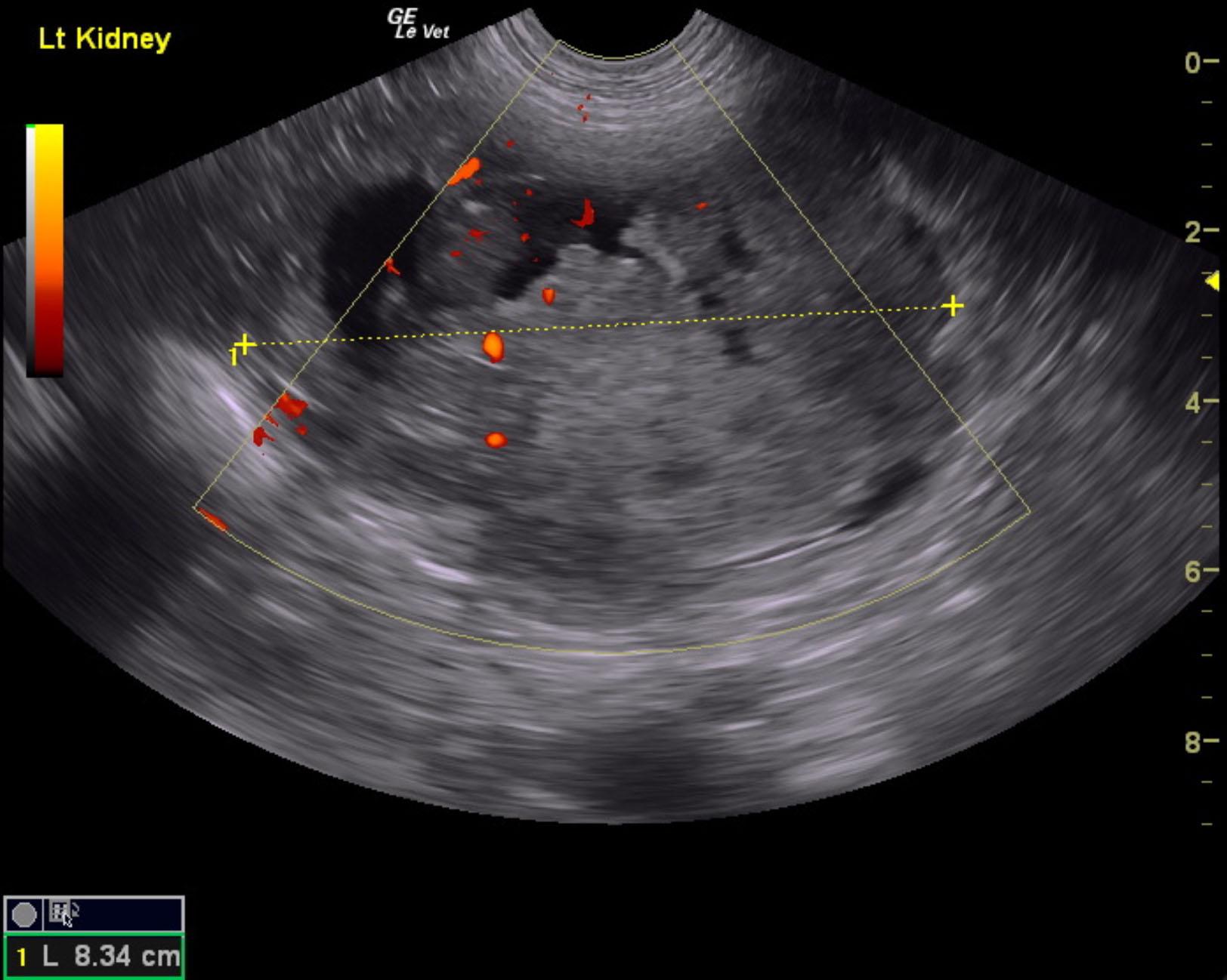
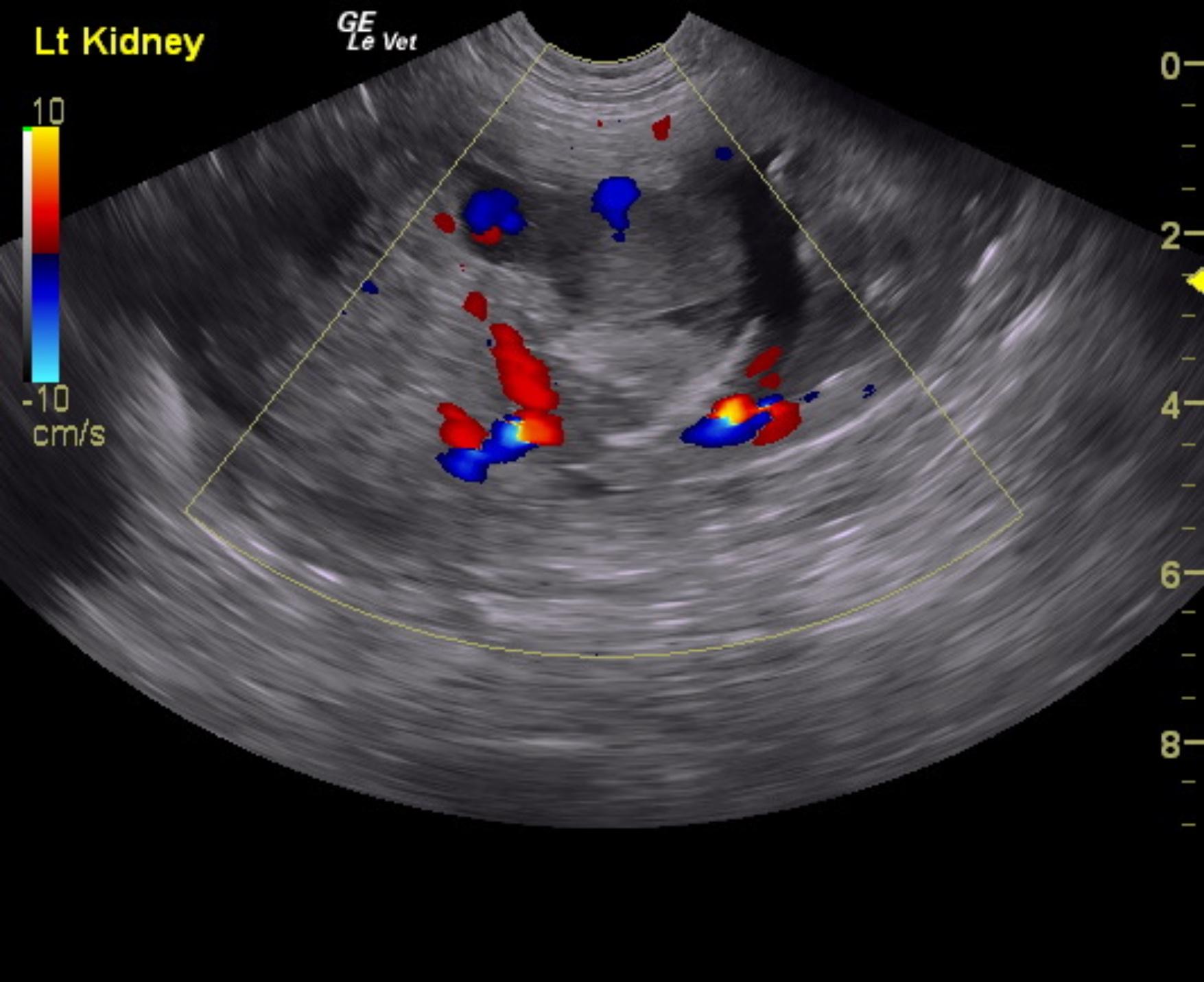
The left kidney in this patient presented a complex, mixed echogenic cystic and disrupted mass measuring 10.4 x 5.1 cm. Power Doppler assessment of the left mass revealed moderate vascularity. Carcinoma is suspected. The left ureter was dilated and deviated owing to the mass effect. The right kidney was unremarkable and normal in size and contour measuring 7.06 cm. Normal color flow was noted.
None
Renal mass – neoplasia, hydronephrosis, hematoma, pyelonephritis, abscess, granuloma
None