A 15-year-old NM Poodle was presented for evaluation of stranguria and pollakiuria. Abnormalities on urinalysis were 1+ blood and 4+ protein.
A 15-year-old NM Poodle was presented for evaluation of stranguria and pollakiuria. Abnormalities on urinalysis were 1+ blood and 4+ protein.
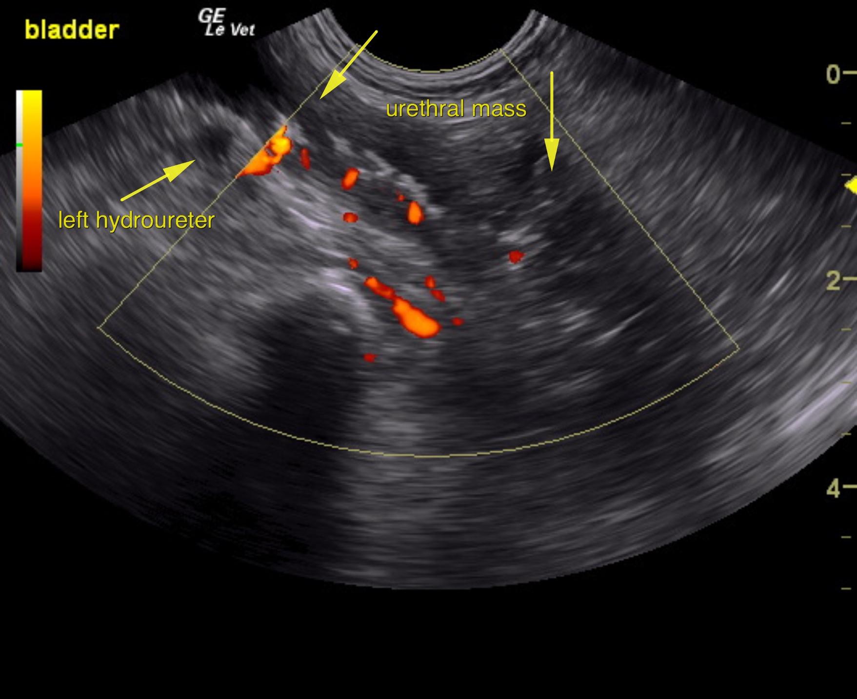
Urethral and prostatic carcinoma pattern with mineralization and mild to moderate left hydroureter with moderate hydronephrosis of the left kidney.
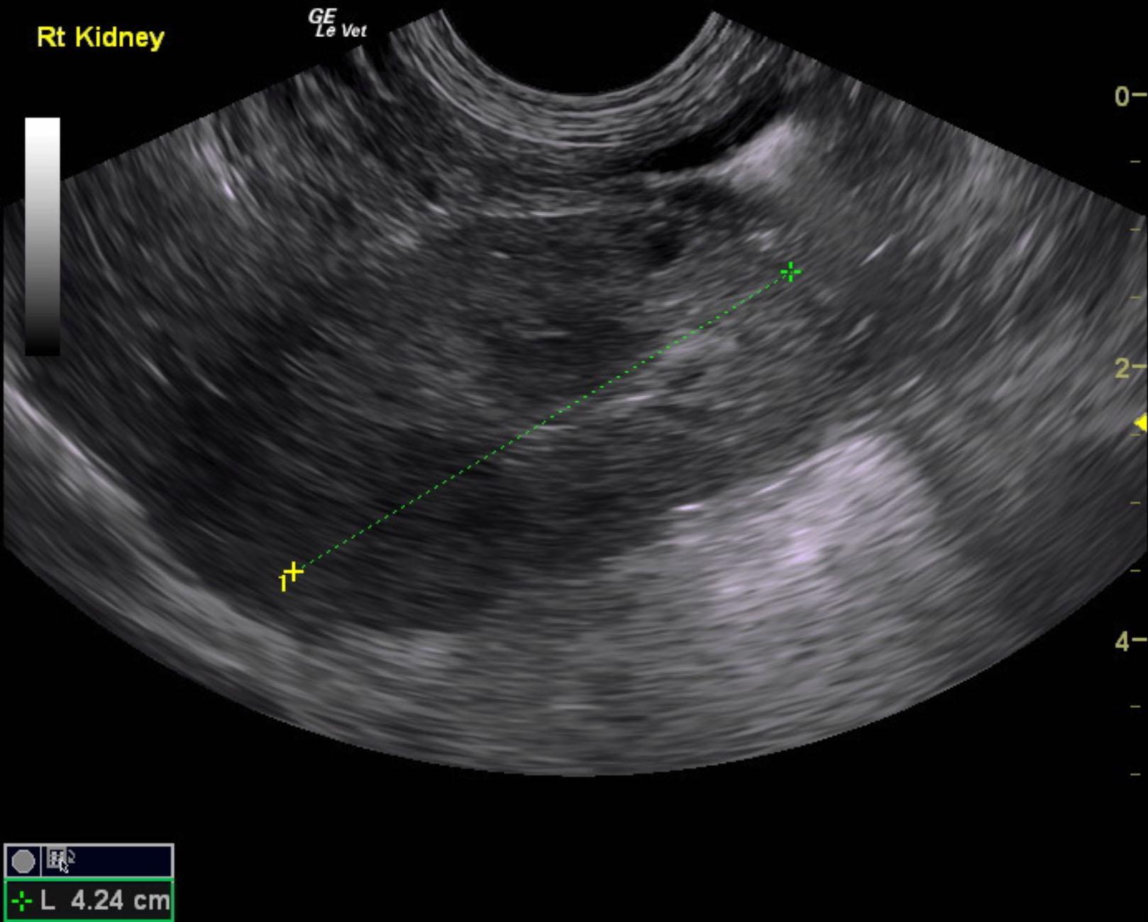
Mild right hydroureter with mild to moderate degenerative right kidney changes.
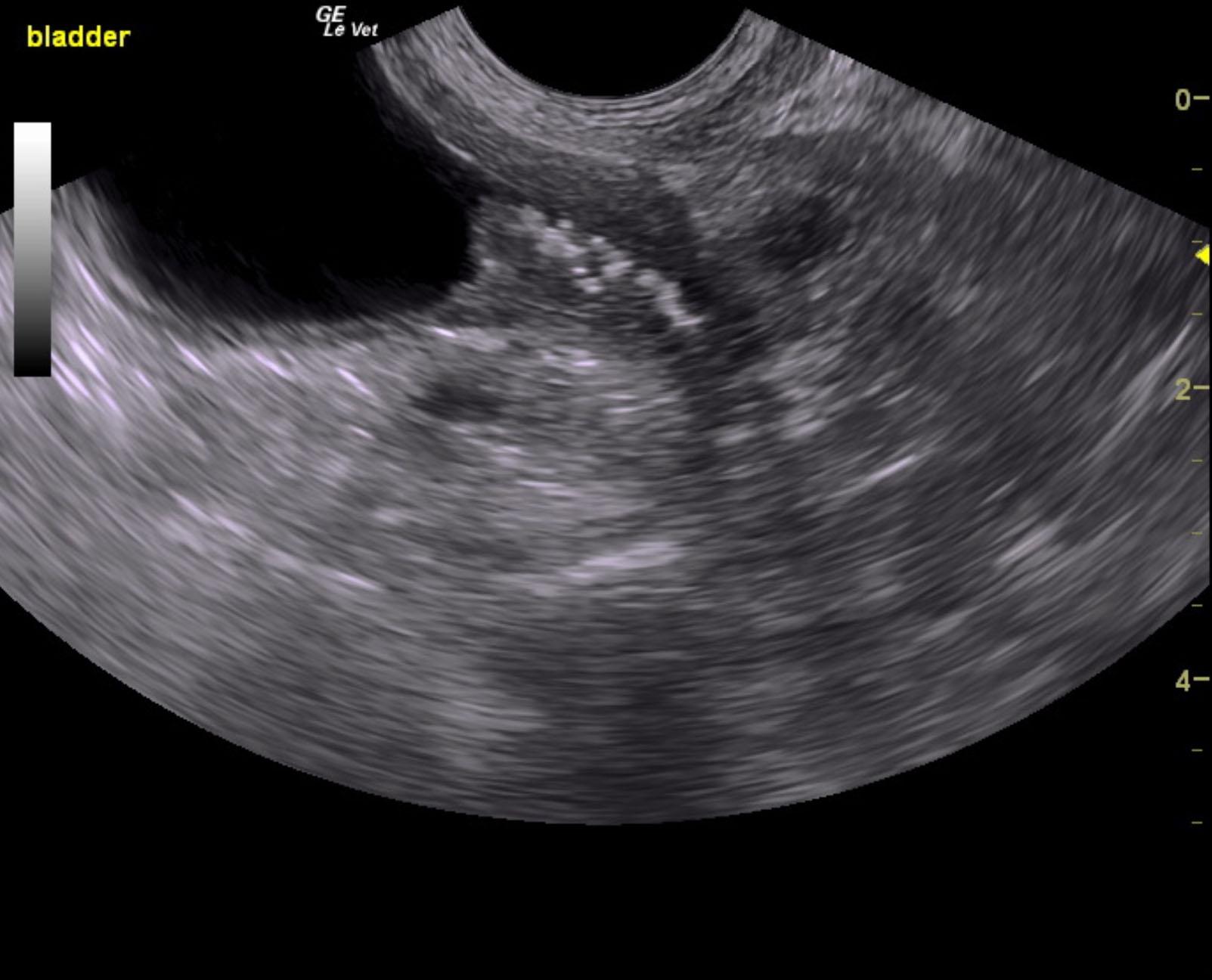
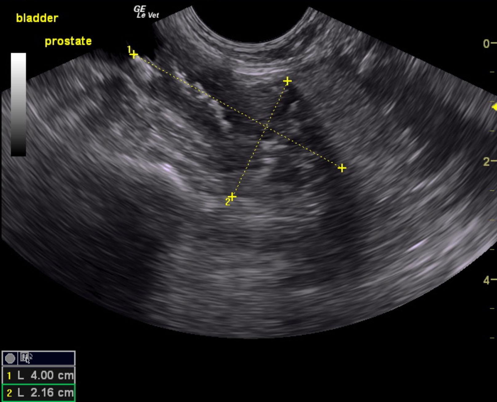
The urinary bladder presented an expansive, mineralizing mass that extended at least 4.0 x 2.16 cm past the cystourethral junction. Obstruction of the left ureter and partial obstruction of the right ureter was noted. The left ureter measured approximately 0.5 cm in width. The mass extends into the region of the prostate completely destroying the prostatic tissue.
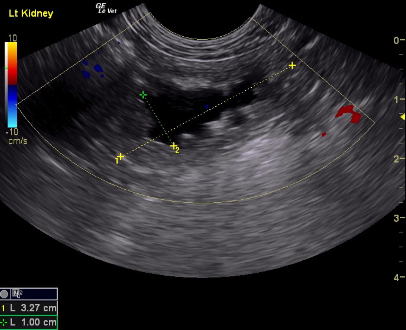
The right kidney presented mild degenerative changes and anechoic cortical cysts. The right kidney revealed loss of corticomedullary definition and hydronephrosis. The right kidney measured 4.24 cm. The left kidney presented moderate hydronephrosis. The left kidney measured 3.27 cm with a 1.0 x 2.0 cm hydronephrotic renal pelvis.
None
Bladder – neoplasia, chronic cystitis, uroliths, polyploid cystitis
Prostate – neoplasia, prostatitis
Urethra – lith, neoplasia
None