A 13-year-old SF Dachshund with a history of frequent UTI and recent bladder stones was presented for evaluation of recent onset stranguria and hematuria. Urinalysis showed SG of 1.015, hematuria, and negative culture
A 13-year-old SF Dachshund with a history of frequent UTI and recent bladder stones was presented for evaluation of recent onset stranguria and hematuria. Urinalysis showed SG of 1.015, hematuria, and negative culture
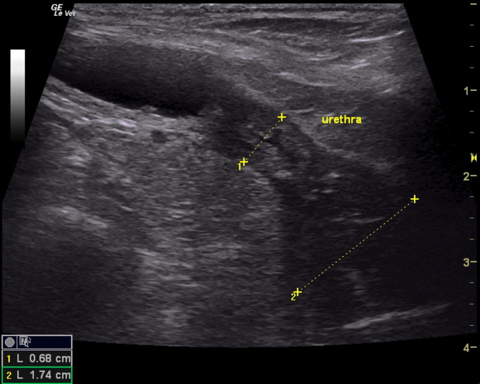
Expansive urethral mass initiating from the cystourethral junction to the deep pelvis.
Focal mineralization. This is strongly suggestive for transitional cell carcinoma. Ultrasound-guided laser ablation would be optimal for this patient. There was no obvious metastatic disease noted. Alternatively stent placement could also be considered, yet the urethra is infiltrated by the mineralizing mass for at least 3.5-4.0 cm caudal from the cystourethral junction. The ureteral papillae were free of evident pathology.
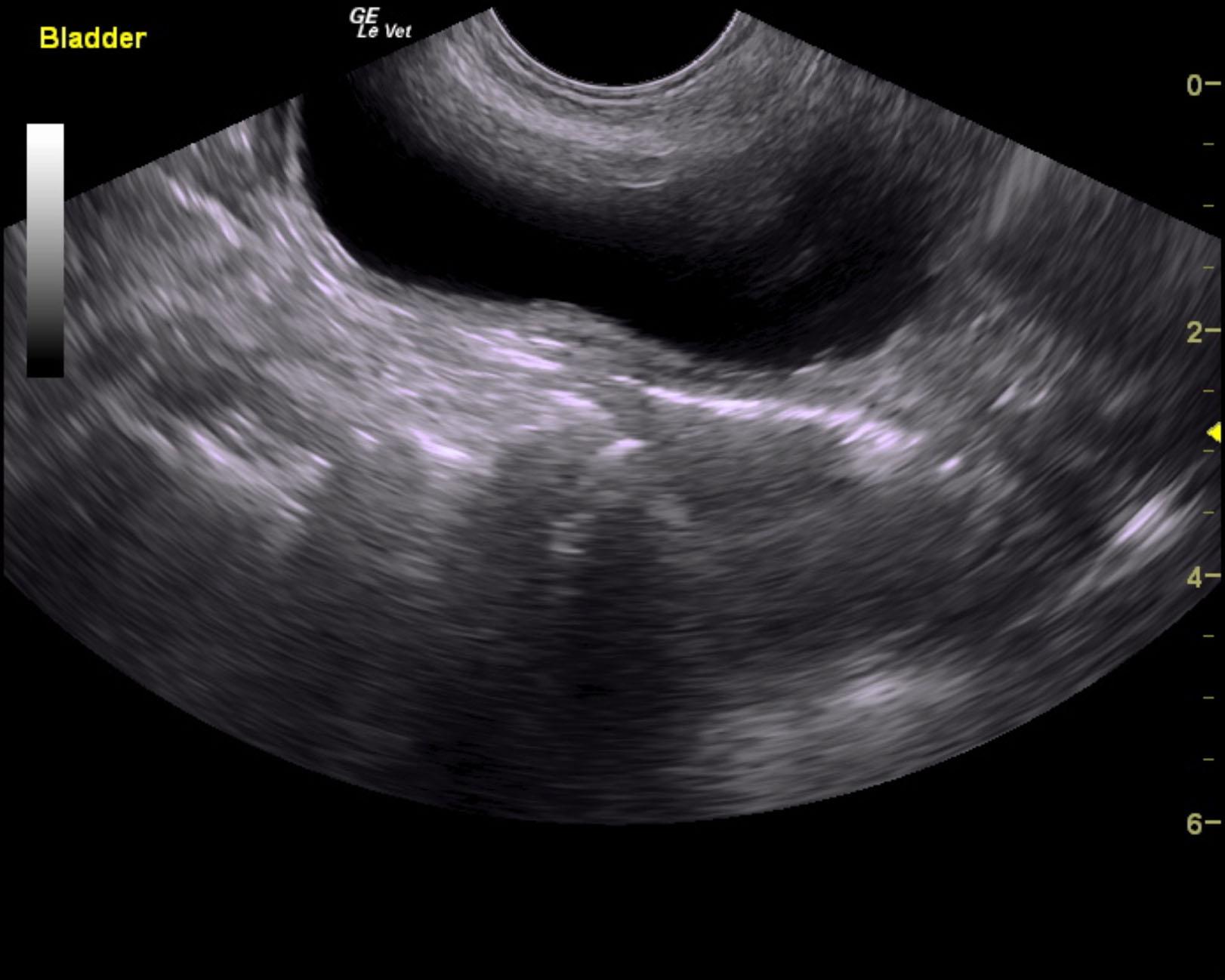
The urethra in this patient presented a mineralizing, expansive mass. This initiated at the cystourethral junction and expanded caudally with pericapsular inflammatory pattern. The cranial portion of the urethral thickening measured 0.68 cm. The caudal portion in the pelvis measured approximately 3.0 cm distal from the cystourethral junction and measured 1.74 cm in width with multiple areas of mineralization. Only minor bladder wall changes were noted, which is consistent with chronic cystitis with a minor potential for spread of the urethral pathology. The bladder, except at the trigone, was unremarkable.
None
Bladder – neoplasia, chronic cystitis, uroliths, polyploid cystitis
Urethra – lith, neoplasia
None