06_00329 Gunner M Congenital renal dysplasia

Case Study

06_00329 Gunner M Congenital renal dysplasia

A 6-month-old male Catahoula was presented for evaluation of renal failure. On physical examination, the face was swollen.


Sonographic Differential Diagnosis

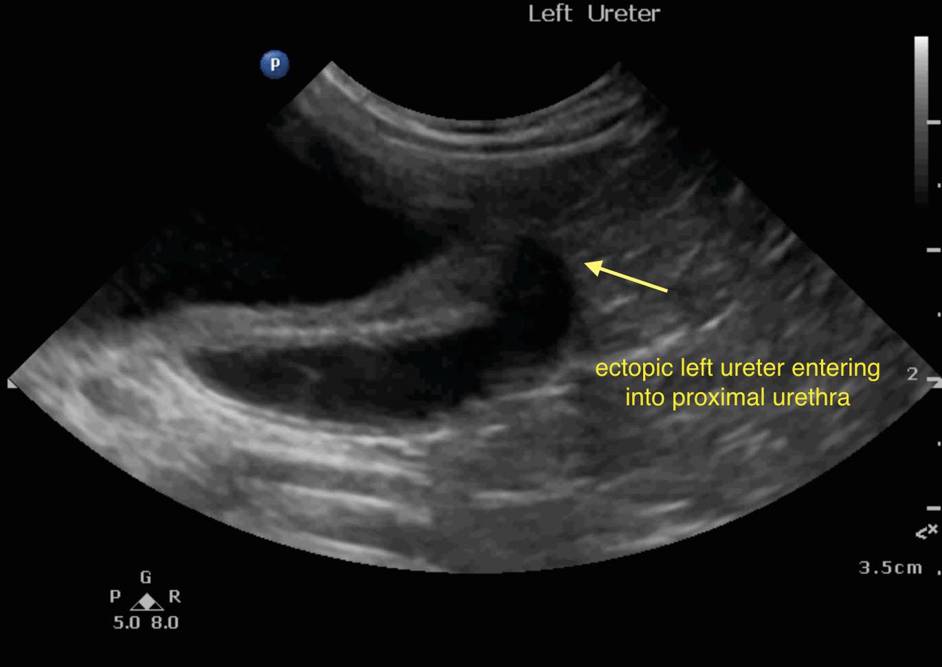
Bilateral renal dysplasia with left ureteral ectopy. Core renal biopsy and ultrasound guided pyelocentesis is warranted for appropriate culture. Renal biopsy to confirm suspicion of primary renal dysplasia.

Image Interpretation

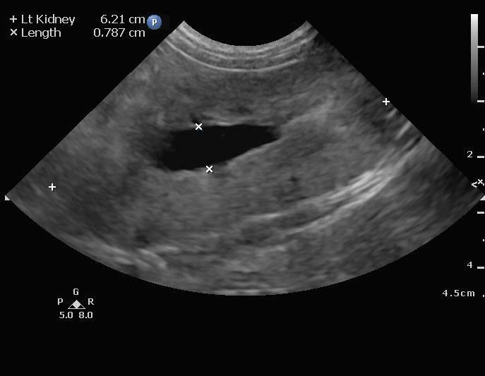
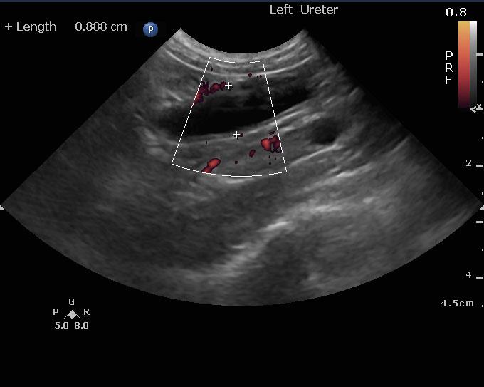
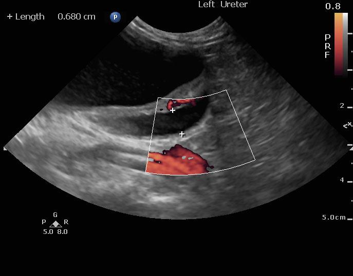
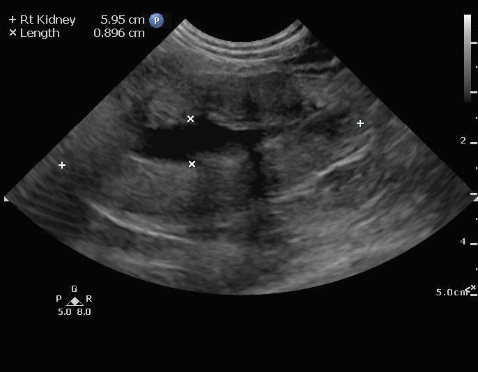
The left kidney in this patient presented dilated renal pelvis, complete lack of corticomedullary structure, thickened irregular cortex. Pyelectasia measured 0.78 cm. Left kidney length 6.21 cm. Right kidney length 5.95 cm with pyelectasia at 0.89 cm similar to the left kidney. Left ureter is ectopic.

DX

Congenital renal dysplasi

Outcome

None

Clinical Differential Diagnosis

Renal – renal dysplasia, pyelonephritis, acute kidney injury (trauma, toxins, hypotension, hypoxia), bacterial nephritis

Sampling

None

Patient Information

Gender : Male, Intact
Species : Canine
Type of Imaging : Ultrasound
Status : Complete

Clinical Signs

  • Swelling

Images

110615_gm_gardens_martinez_20151106_121829_0007110615_gm_gardens_martinez_20151106_121829_0010110615_gm_gardens_martinez_20151106_121829_0012screen_shot_2015-11-07_at_7110615_gm_gardens_martinez_20151106_121829_0022

Clinical Signs

  • Swelling
Skip to content