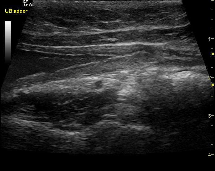
A 5-year-old SF DSH was presented for evaluation of recurrent hematuria
Sonographic Differential Diagnosis
Renal and ureteral calculi.
Image Interpretation
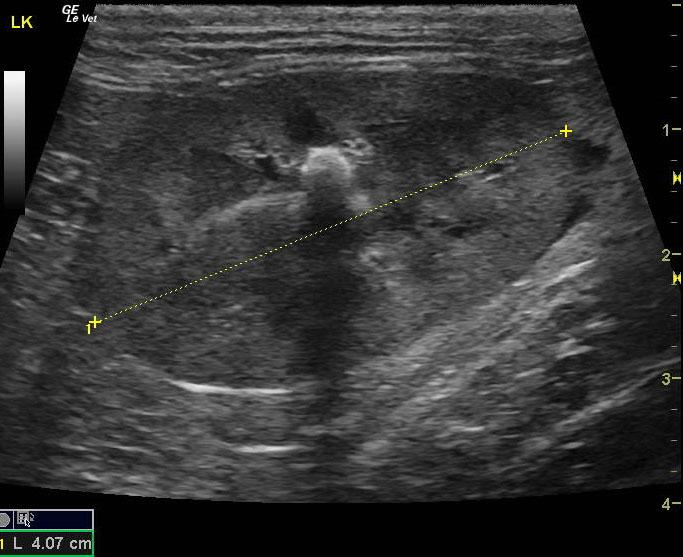
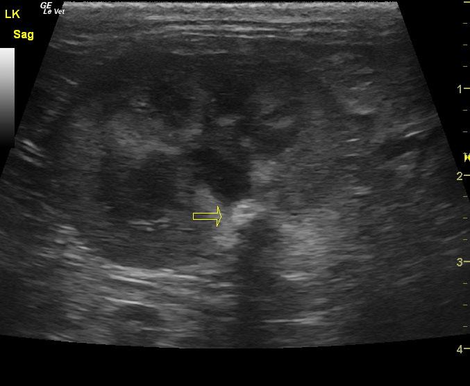
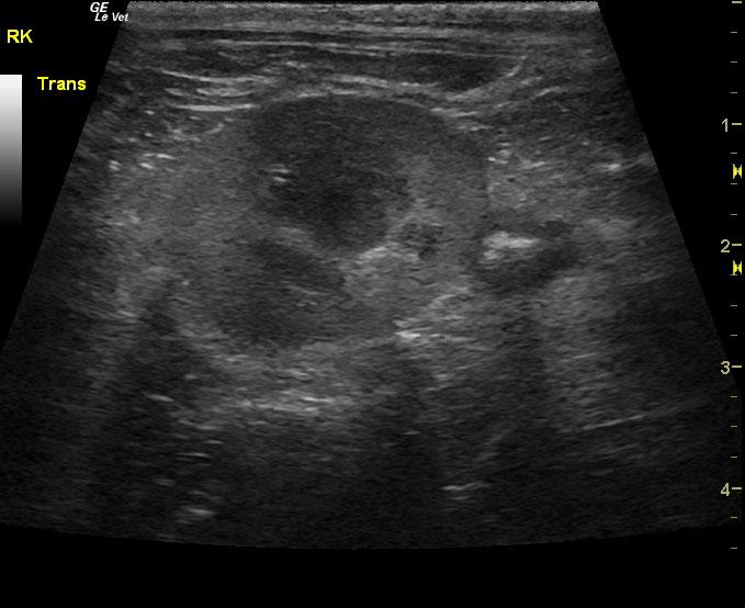
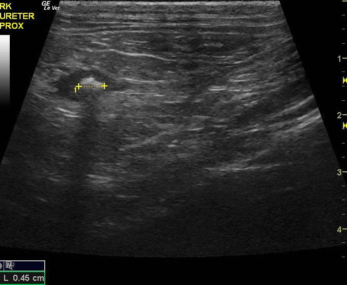
The left kidney presented a 0.52 cm calculus at the corticomedullary junction. It is about to enter into the renal pelvis. A second calculus was noted in the left kidney and measured 0.4 cm. This calculus dilated the pelvis and entered into the proximal left ureter. This is partially obstructive and large enough to be interventionally removed. The left ureter presented a slight calculus that measured 0.18 cm with hydroureter measuring 0.5 cm near the left renal pelvis. The left kidney revealed slight pyelectasia of 0.5 x 1.0 cm. Pericapsular inflammatory pattern and fluid pattern was noted around the left kidney. The right ureter presented a calculus in the proximal right ureter measuring 0.5 cm. The right kidney was uniform and measured 3.85 cm. The pelvic urethra was slightly thickened, yet uniform and imaged 3.0 cm beyond the cystourethral junction.