06_00305 Sadie B Mineralized urethral mass

Case Study

06_00305 Sadie B Mineralized urethral mass

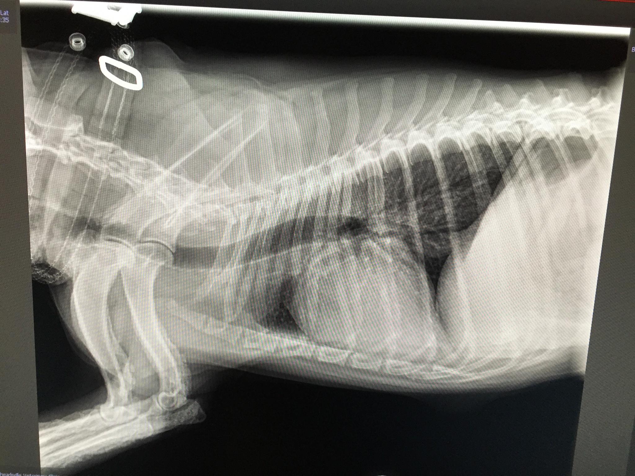
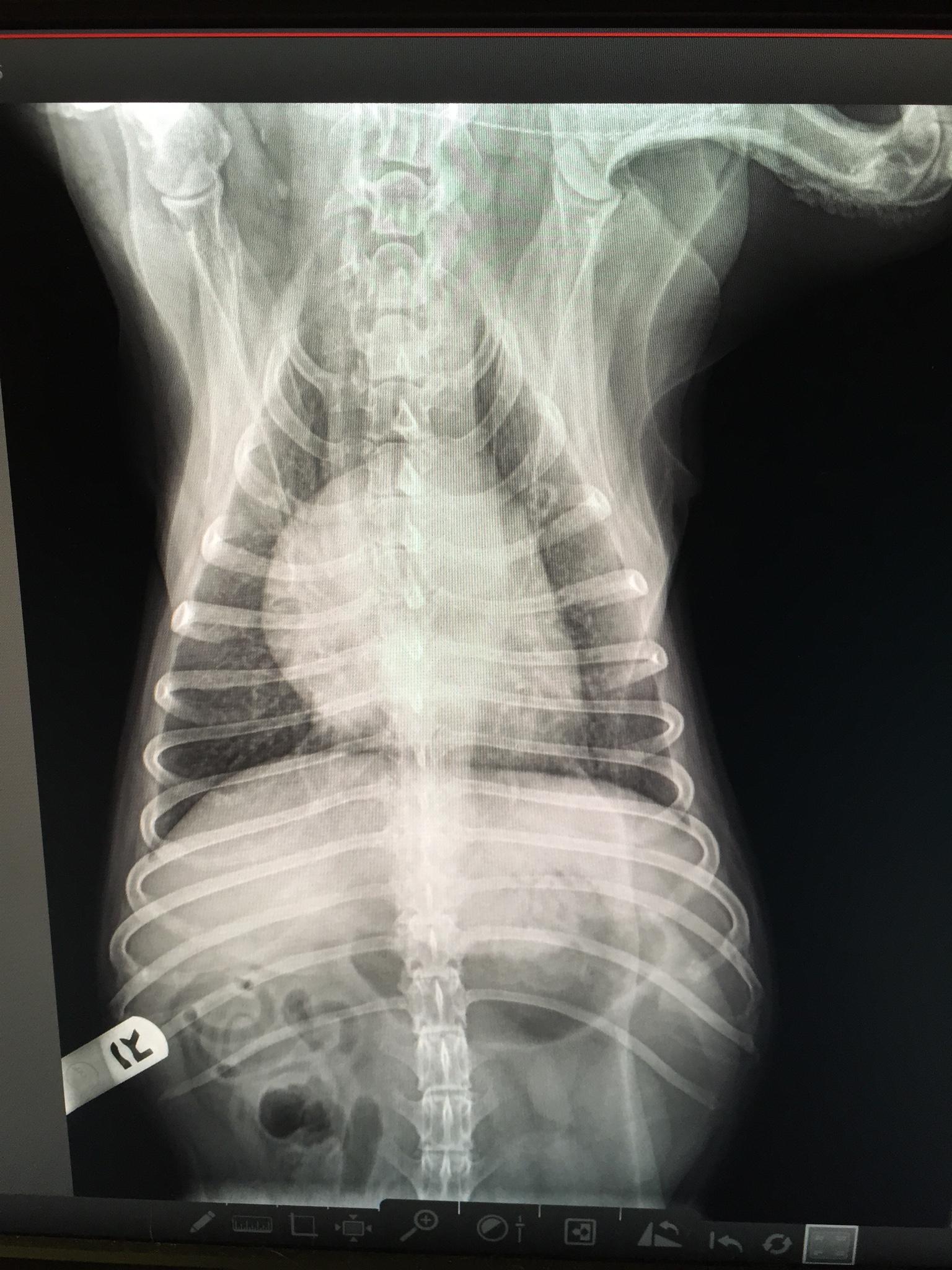
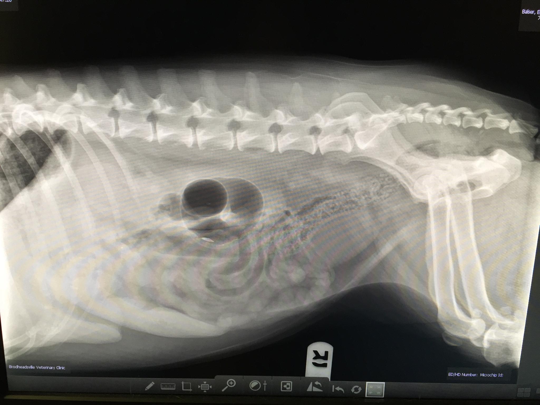
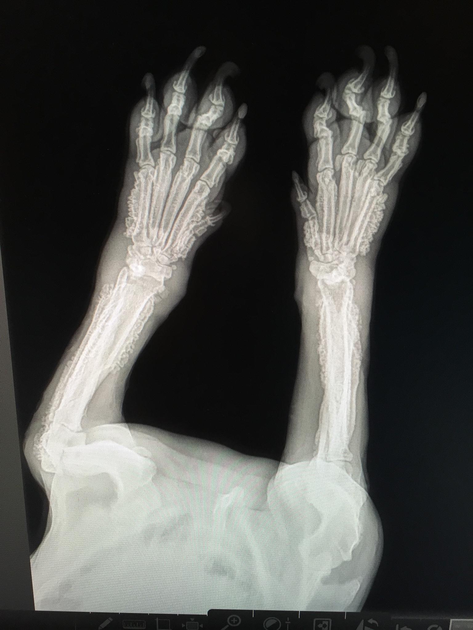
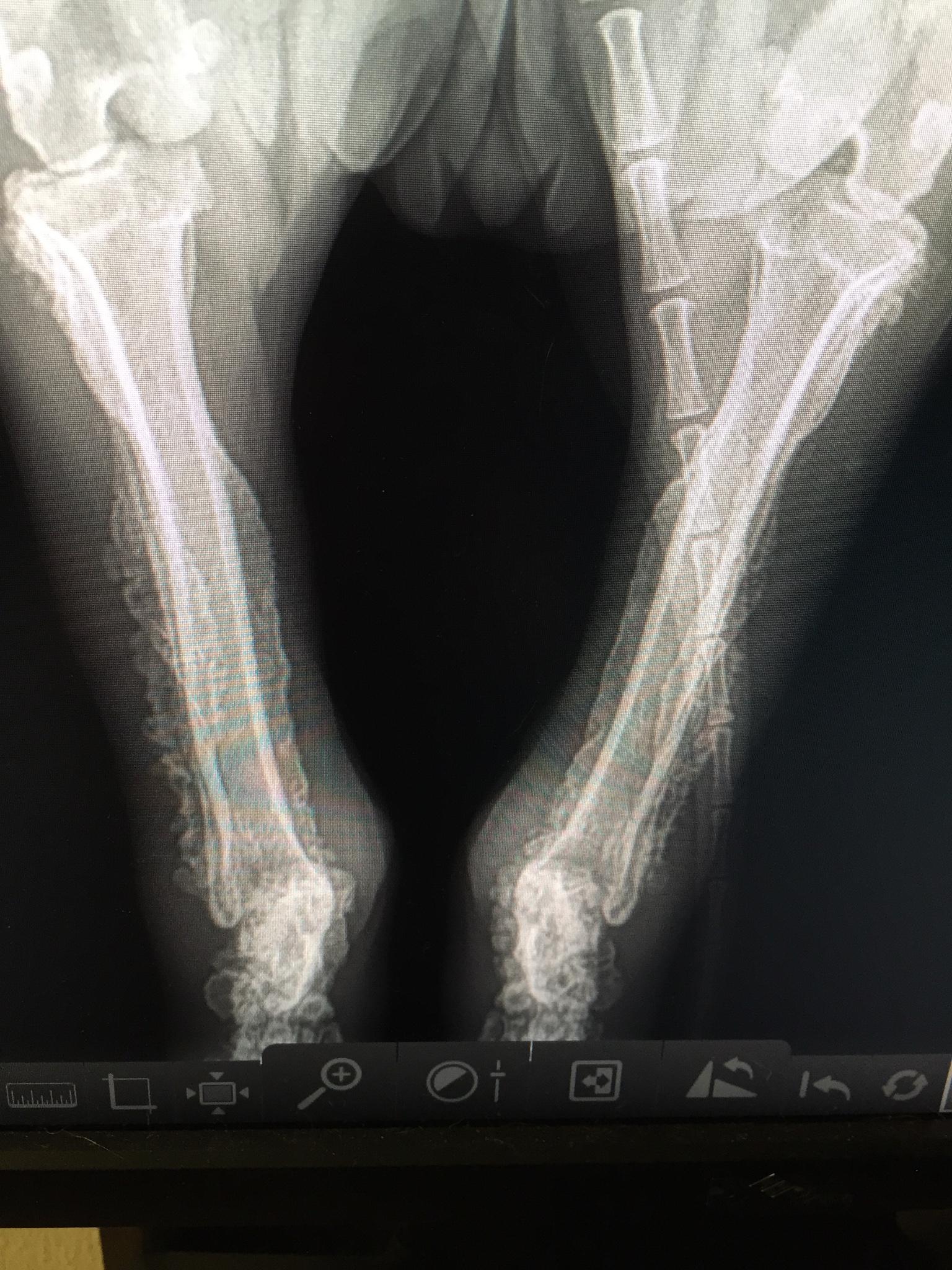
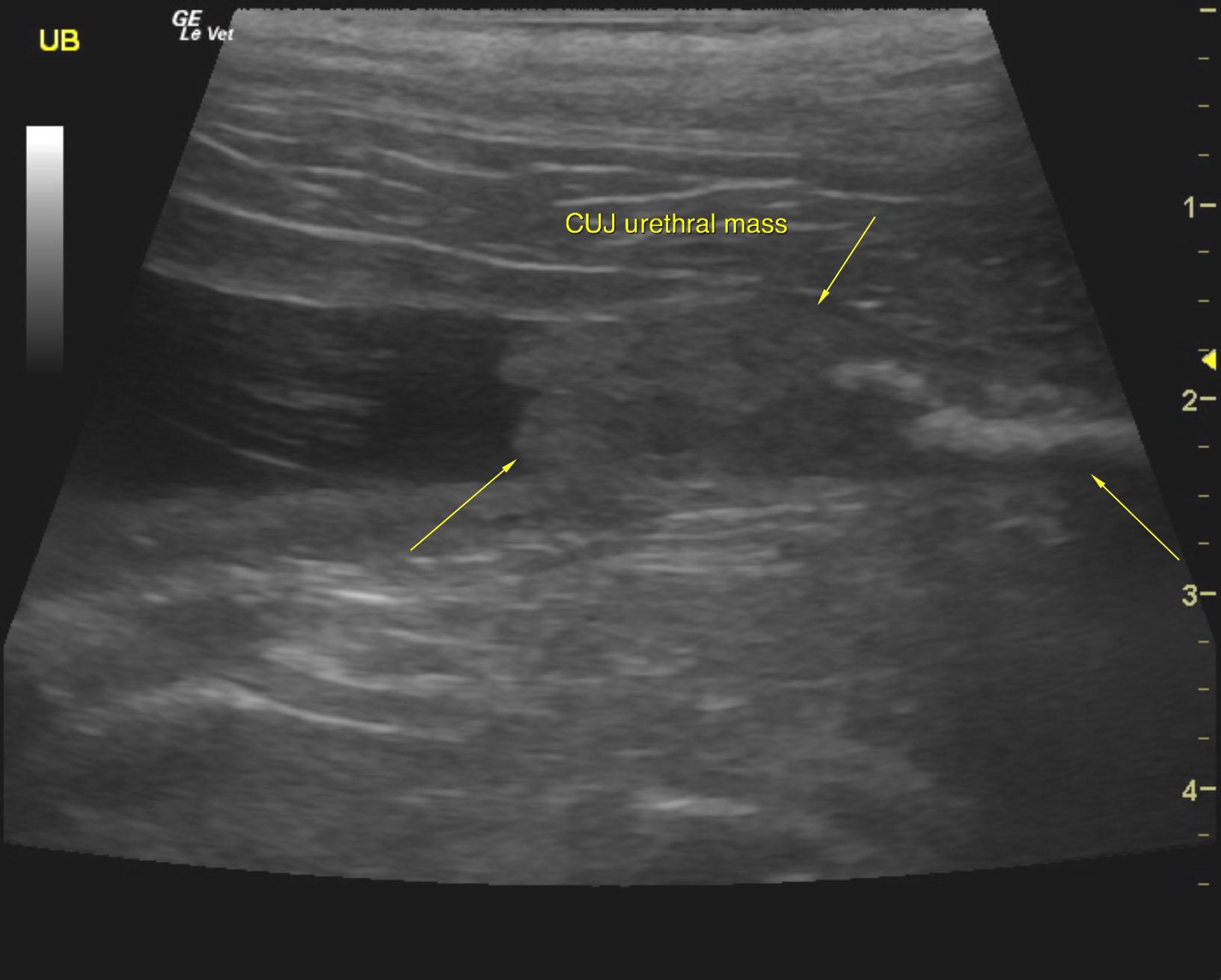
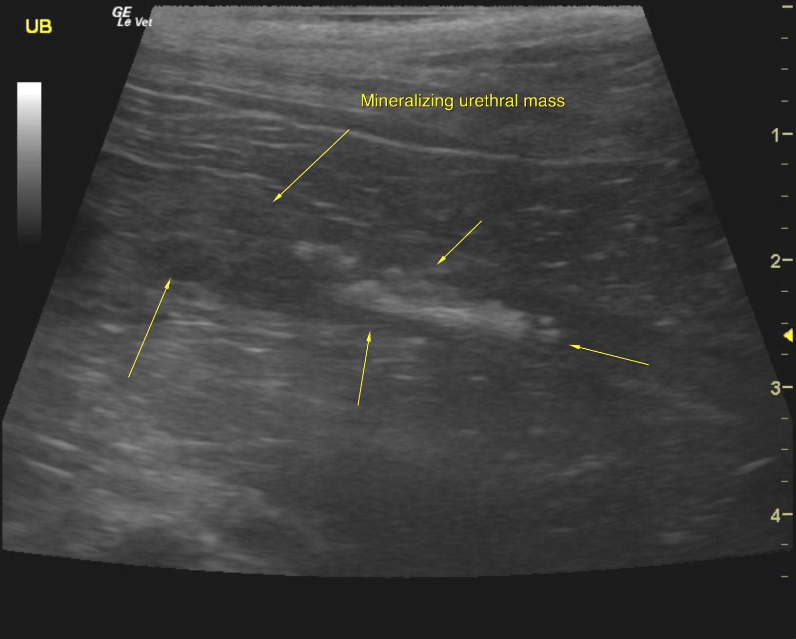
A 10-year-old SF Beagle was presented for evaluation of dysuria and stranguria. Urinalysis showed specific gravity 1.015, 1+ protein, hematuria, and 4+ epithelial cells. Serum biochemistry was normal. Hypertrophic osteopathy was evident on survey radiographs.


Sonographic Differential Diagnosis

Urethral and cystourethral junction mass. Referral for ultrasound guided laser ablation procedure would be ideal in this case given the location of the mass. However, clinical management of the hypertrophic osteopathy may be related or possibly completely unrelated to the urethral mass itself.

Image Interpretation

Pelvic urethra revealed a mineralizing mass. The urethral mass occupied the cystourethral junction. Some minor apical polypoid changes were noted also in the urinary bladder.

DX

Mineralizing urethral mass

Outcome

None

Comments

Liz, I don’t know if all the rads show the HO. Obviously, delete or recaption any that don’t. 

Clinical Differential Diagnosis

Bladder – neoplasia, uroliths, bacterial cystitis, polyploid cystitis
Urethra – neoplasia, granulomatous urethritis, lith
Hypertrophic osteopathy – neoplasia/abscess/granuloma within abdomen or thorax

Sampling

None

UA Specific Gravity Range

1.015

Patient Information

Gender : Female, Spayed
Species : Canine
Type of Imaging : Ultrasound
Status : Complete

Clinical Signs

  • Dysuria
  • Stranguria

Images

img_4064img_4066img_4065img_4067img_4068sadie_urethral_mass_cujsadie_urethral_mass

Clinical Signs

  • Dysuria
  • Stranguria

Urinalysi

  • Blood Present
  • Epithelial Cells Present
  • Protein Present
  • Specific Gravity Low
Skip to content