06_00304 Pandora C Hydronephrosis, acute on chronic nephritis, renal and bladder calculi

Case Study

06_00304 Pandora C Hydronephrosis, acute on chronic nephritis, renal and bladder calculi

 A 10 year-old SF DSH with a history of renoliths was presented for evaluation of a week’s duration of vomiting, inappetence, and lethargy. Isosthenuria was present on urinalysis.  Abnormalities on CBC and serum biochemistry were anemia, hyperkalemia, and azotemia. Survey radiographs showed bladder and renal calculi.

Sonographic Differential Diagnosis

Moderate hydronephrosis and acute on chronic nephritis in the left kidney.
Chronic, mild hydronephrosis/pyelectasia in the right kidney with moderate to severe dystrophic changes.
Renal and bladder calculi. This patient has likely been passing calculi periodically with a recent acute event. Secondary ureteral strictures appear to be an issue.

Image Interpretation

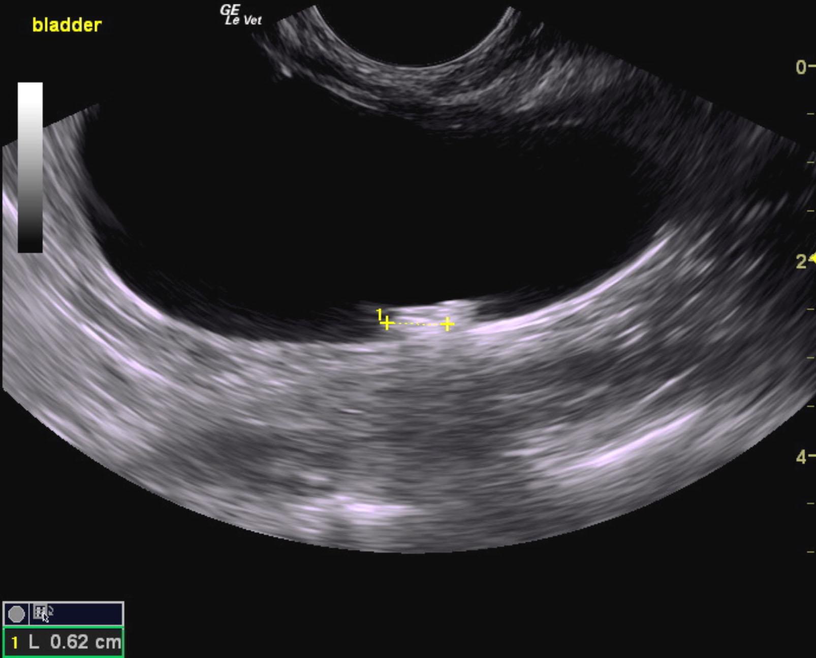
The urinary bladder appeared unremarkable. Bladder calculi were noted with an accumulation of approximately 1.0 cm. The largest calculus measured 0.62 cm.
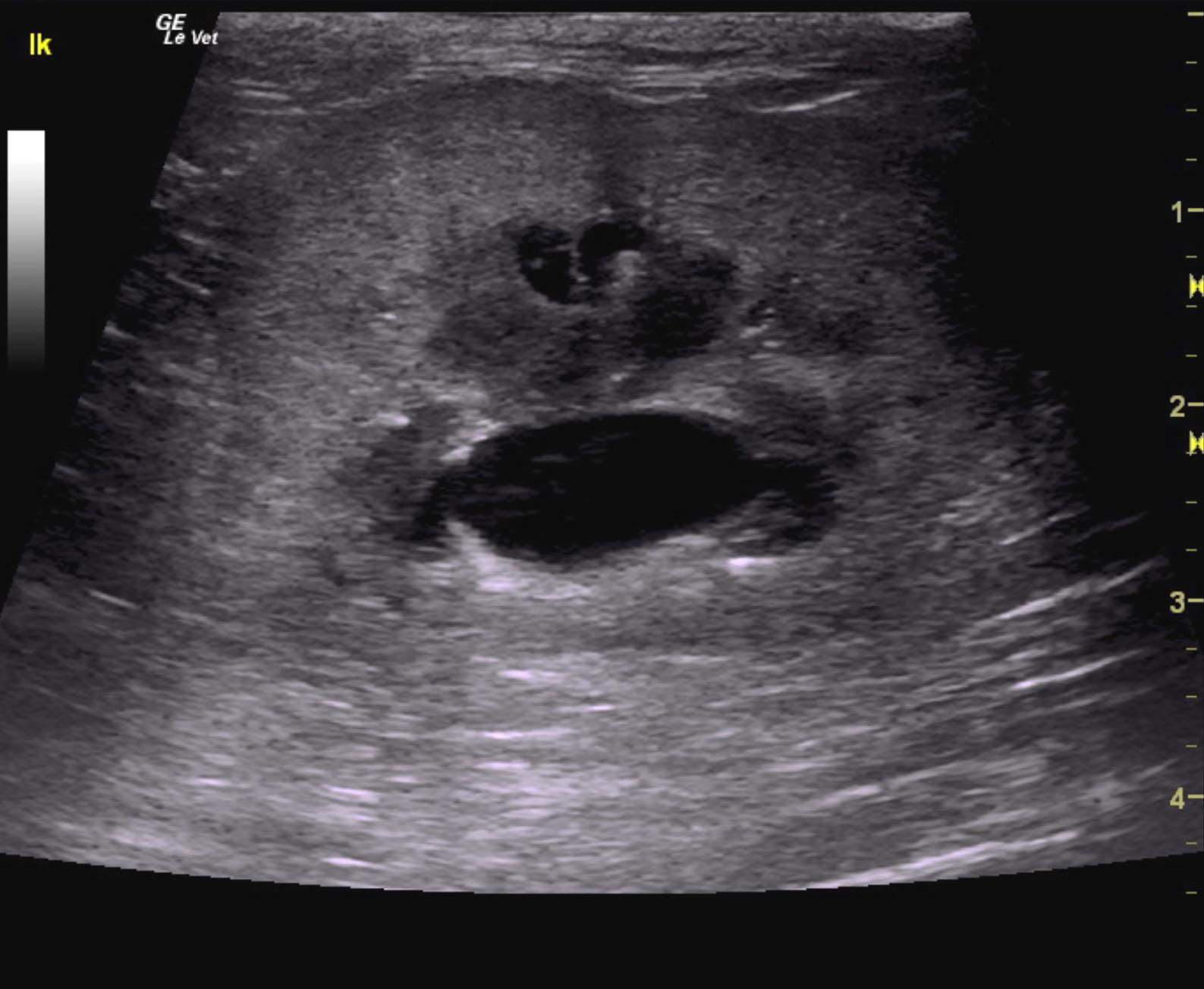
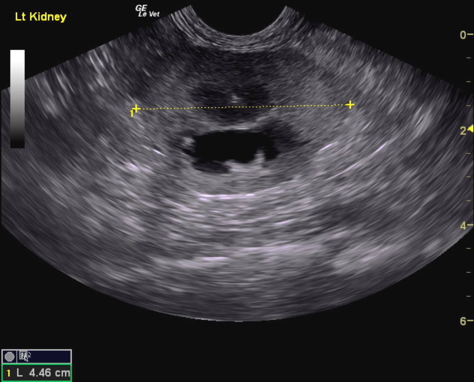
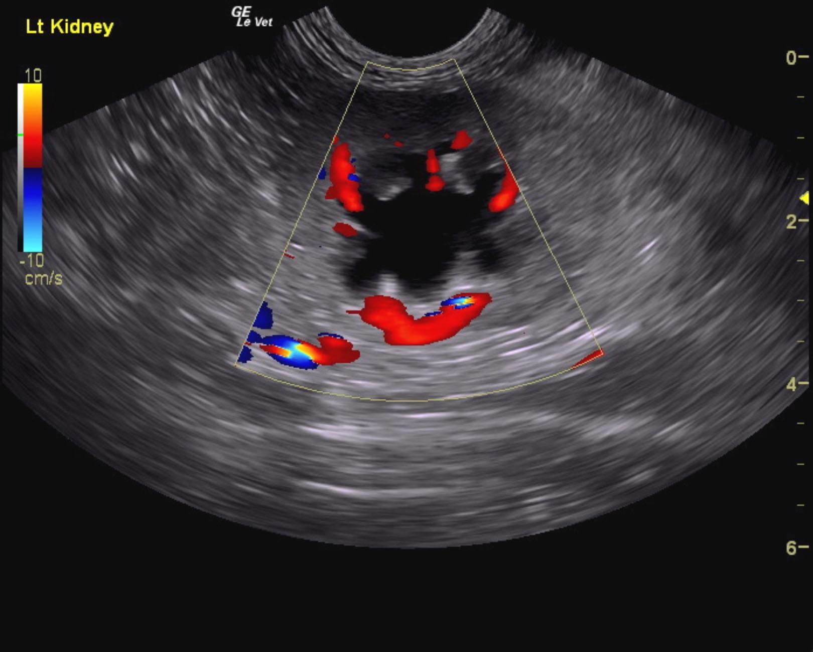
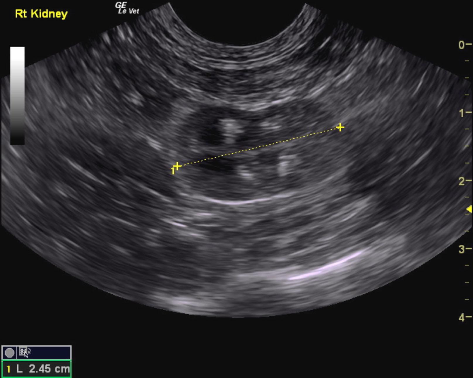
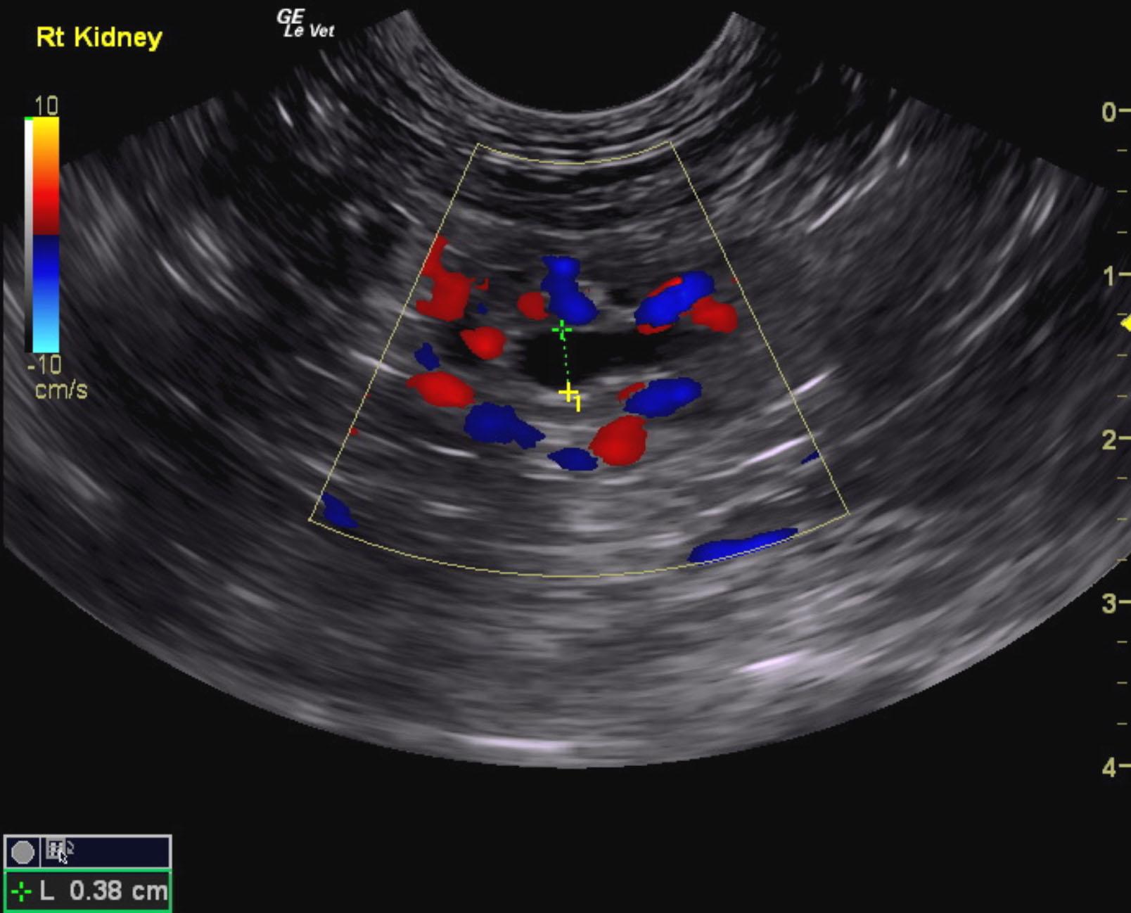
The kidneys in this patient presented moderate to severe dystrophic changes, irregular contour, cortical infarcts and remodeling were noted. The left kidney presented moderate hydronephrosis with interstitial nephrosis pattern. Swollen contour and periserosal inflammatory pattern was noted. Color flow assessment of the renal cortex appeared adequate. However, significant disruption of architecture was noted. The right kidney revealed pyelectasia that measured 0.38 cm with no obstructive calculi at this time. The right kidney was subnormal in size and measured 2.45 cm. The left kidney measured 4.46 cm. The left kidney also revealed pyelectasia that measured 0.73 x 1.5 cm. Slight, pericapsular fluid accumulation was noted near the left kidney. This is consistent with acute nephritis owing to hydronephrosis. The left proximal ureter revealed either a stricture or calculus that measured approximately 1.0 cm from the left renal pelvis. However, this is more consistent with stricture.

DX

Hydronephrosis, acute on chronic nephritis, renal and bladder calculi

Outcome

None

Clinical Differential Diagnosis

Renal – renoliths, pyelonephritis, obstructive uropathy, acute kidney injury superimposed on chronic kidney disease, Leptospirosis
Bladder uroliths

Sampling

None

Patient Information

Gender : Female, Spayed
Species : Feline
Type of Imaging : Ultrasound
Status : Complete

Clinical Signs

  • Anorexia
  • Lethargy
  • Vomiting

Images

pandora_cushing_renal_stone_passage_renal_failure_ub_stonespandora_cushing_renal_stone_passage_renal_failure_lk_2pandora_cushing_renal_stone_passage_renal_failure_lk_3pandora_cushing_renal_stone_passage_renal_failure_lkpandora_cushing_renal_stone_passage_renal_failure_rkpandora_cushing_renal_stone_passage_renal_failure

Blood Chemistry

  • Azotemia
  • Potassium, High

CBC

  • RBC, Low

Clinical Signs

  • Anorexia
  • Lethargy
  • Vomiting

Urinalysi

  • Isosthenuria Present
Skip to content