A 6-year-old SF Rhodesian Ridgeback was presented for evaluation of dysuria. Urinalysis showed normal SG (1.031), proteinuria, and the presence of red and white blood cells.
A 6-year-old SF Rhodesian Ridgeback was presented for evaluation of dysuria. Urinalysis showed normal SG (1.031), proteinuria, and the presence of red and white blood cells.
Urethral and bladder mass with iliac lymph node mass and regional iliac lymphadenopathy. This is strongly suggestive for metastatic disease. Palliative therapy with ultrasound-guided laser ablation could be considered; however, given the iliac metastatic pattern the utility is dubious. Oncology consultation is recommended.
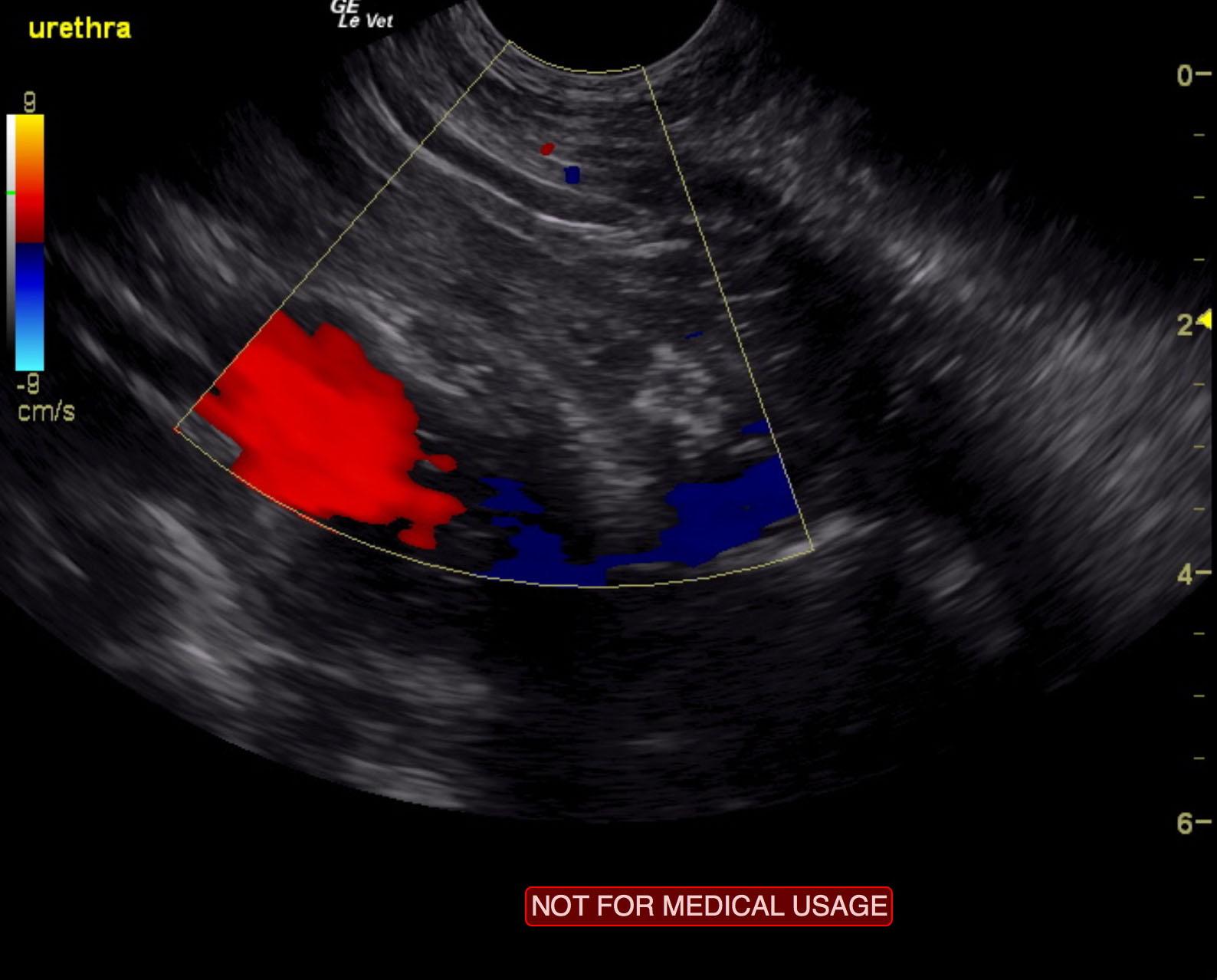
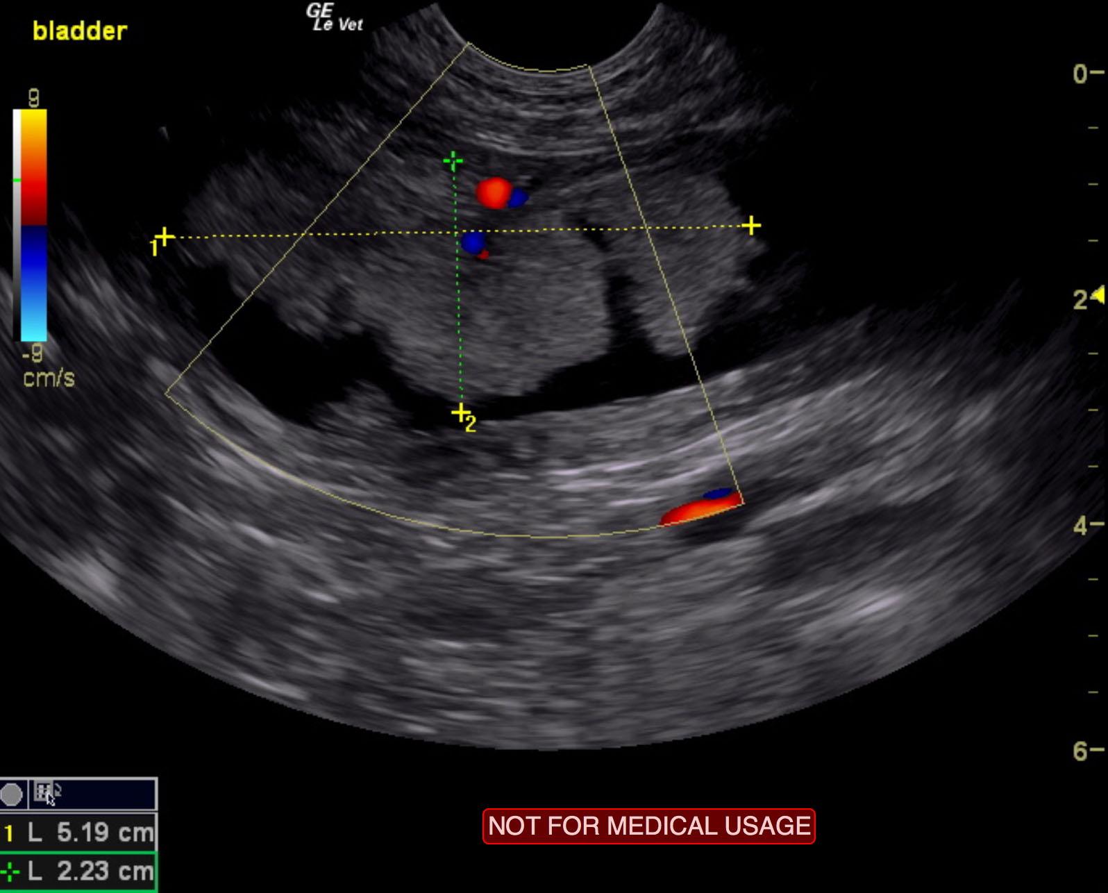
The pelvic urethra revealed a mineralizing mass that measured 1.4 cm in width and extended to at least 4.0 cm caudal from the cystourethral junction. The primary bladder mass measured 5.19 x 2.23 cm. The bladder mass appeared significantly vascular.
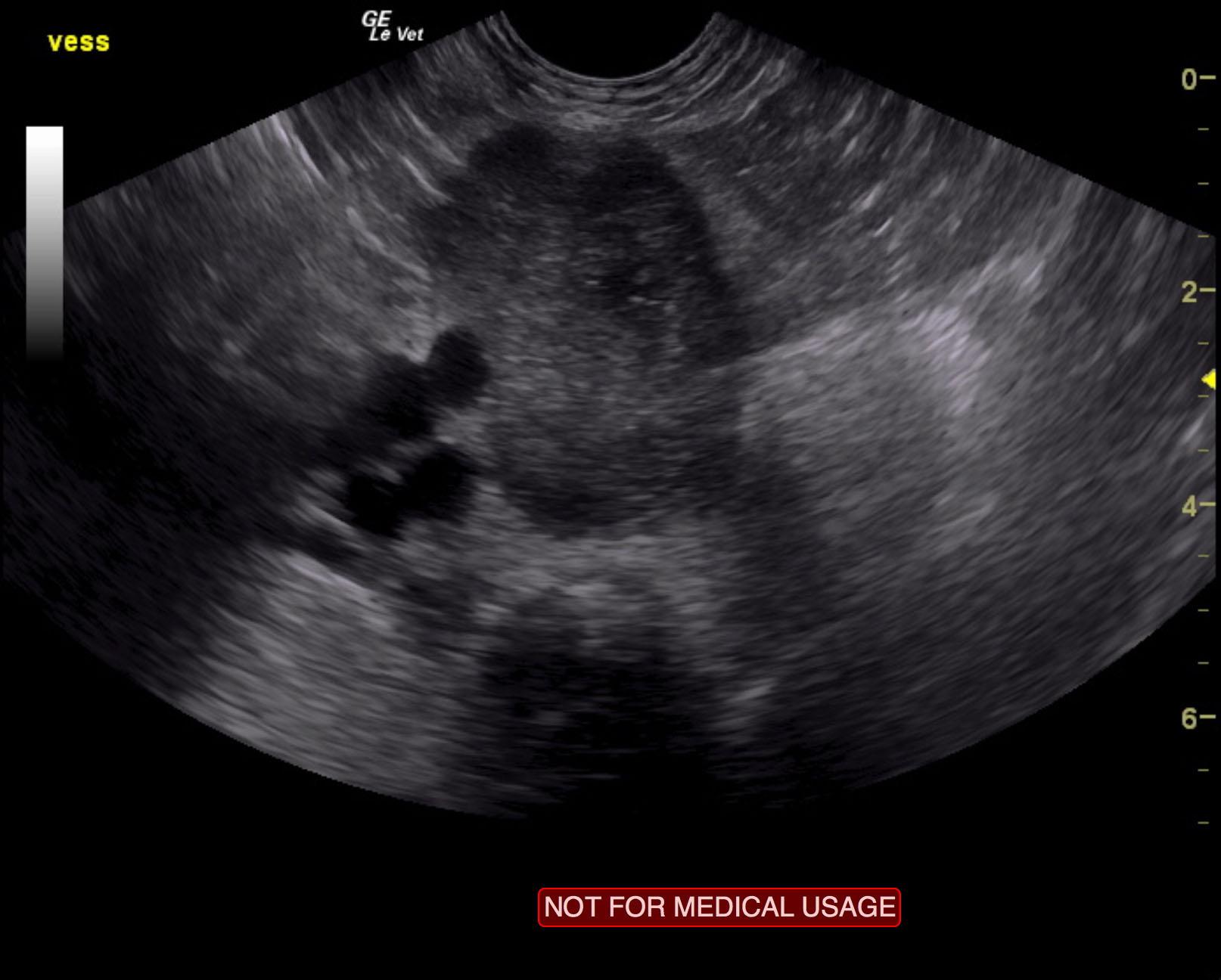
Iliac lymph nodes were mildly enlarged and measured 0.57 cm. A separate lymph node mass was noted and measured 6.4 x 3.98 cm. This is consistent with metastatic disease. Pericapsular inflammatory pattern was noted around the lymph nodes.
None
Urinary bladder – chronic bacterial cystitis, urolith, neoplasia, granulomatous cystitis, polyploid cystitis
Urethra – urolith, neoplasia, granulomatous urethritis
None