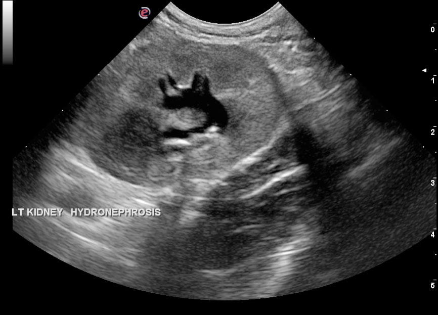
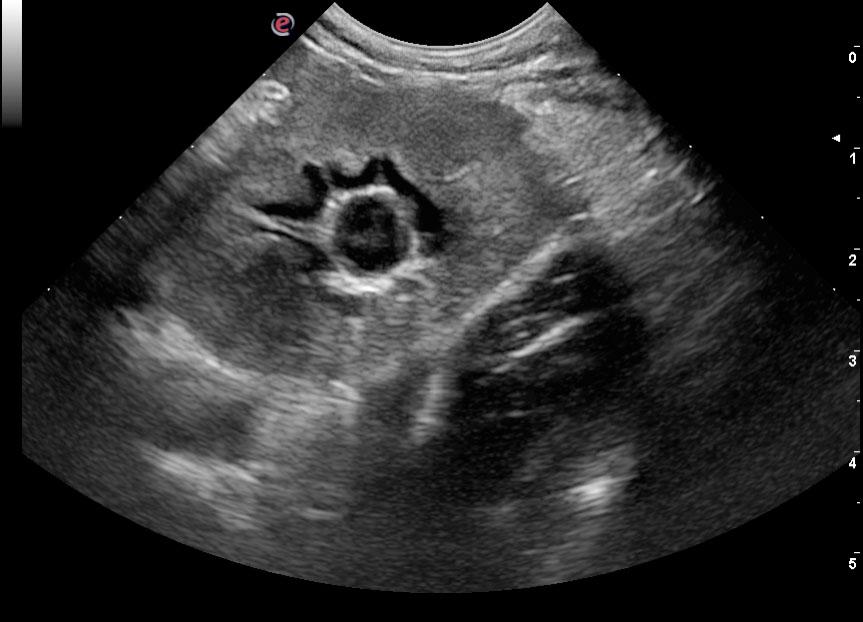
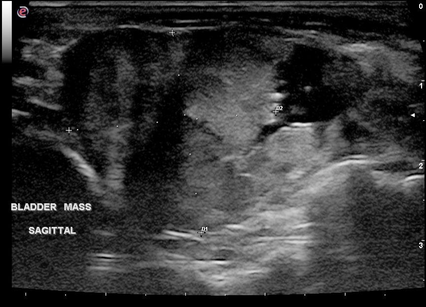
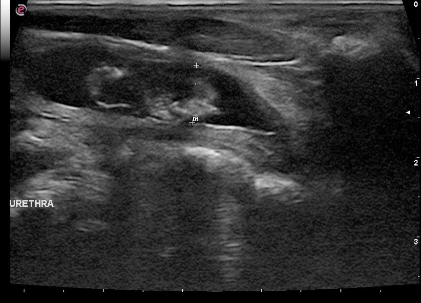
A 10-year-old SF DSH was presented for evaluation of stranguria and hematuria, attributed to chronic suspected urinary tract infection. Additional history was weight loss. On physical examination, a firm irregular easily expressible bladder and hematuria was evident. Hematuria and pyuria was present on urinalysis. CBC showed neutrophilia whereas serum biochemistry was normal and T4 in the grey zone. On survey radiographs, a soft tissue structure within the bladder was evident.
A 10-year-old SF DSH was presented for evaluation of stranguria and hematuria, attributed to chronic suspected urinary tract infection. Additional history was weight loss. On physical examination, a firm irregular easily expressible bladder and hematuria was evident. Hematuria and pyuria was present on urinalysis. CBC showed neutrophilia whereas serum biochemistry was normal and T4 in the grey zone. On survey radiographs, a soft tissue structure within the bladder was evident.