An 11-year-old SF cat was presented for evaluation of a possible caudal abdominal mass. Blood work was within normal limits.
An 11-year-old SF cat was presented for evaluation of a possible caudal abdominal mass. Blood work was within normal limits.
Case Study
Pelvic ectopic kidney in an 11 year old FS cat
Sonographic Differential Diagnosis
Pelvic positioning of the right kidney, ectopic position. Minor, chronic degenerative changes to the left kidney. Potential concurrent urinary tract infection is possible and urinalysis would be indicated. Urine culture and sensitivity would be warranted, as well as blood pressure measurements. There was no evidence of neoplasia noted.
Image Interpretation
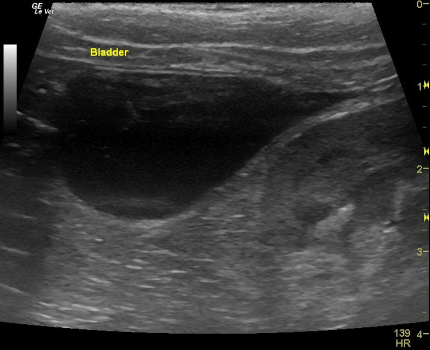
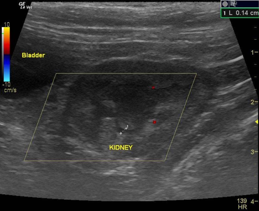
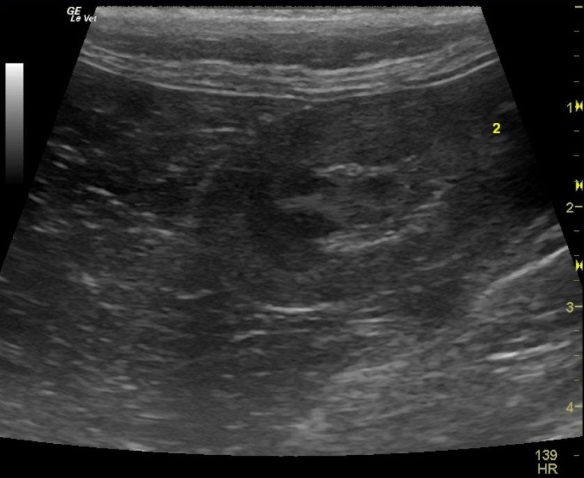
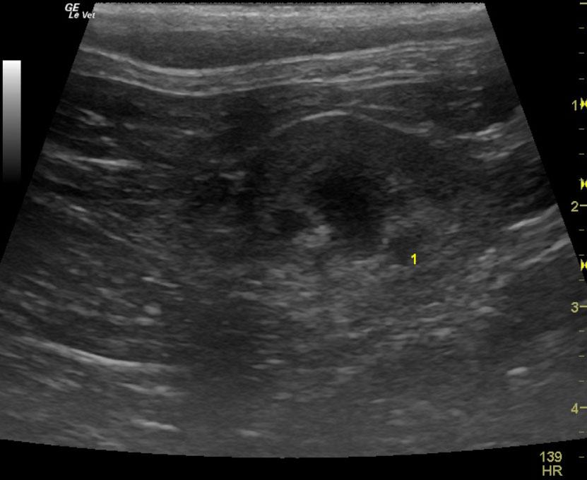
The urinary bladder, trigone and pelvic urethra presented normal wall thicknesses with anechoic urine and normal tone. No uroliths or sediment were visualized. No evidence of inflammatory or neoplastic changes were noted. The ureters were not visible and considered normal. The left kidney was uniform and measured 3.32 cm with minor, medullary rim sign. The right kidney was displaced caudally in this patient with dystrophic changes and minor pyelectasia. It was in ectopic position. This is an infrequent anomaly in cats. The area of the right kidney was free of evident pathology.
DX
Ectopic right kidney
Outcome
No further outcome at this time.
Comments
I invite you to see August 2011 case of the month on www.sonopath.com for a description of this and other positional anomalies in cats.
Clinical Differential Diagnosis
Abdominal mass – neoplasia, granuloma, abscess of kidney, lymph nodes, intestine, bladder, or mesentery, hydronephrosis, peri-nephric cyst.
Sampling
None
Video
Patient Information
Patient Name :
Lelo F
Gender :
Female, Spayed
Species :
Feline
Type of Imaging : Ultrasound
Status :
Complete
Liz Wuz Here :
Yes
Code :
06_00118
Exam Finding
- Palpable mass
Images