05_00255 Maggie D Pancreatitis

Case Study

05_00255 Maggie D Pancreatitis

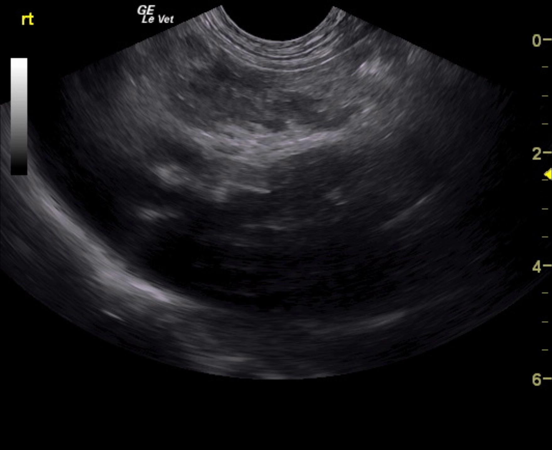
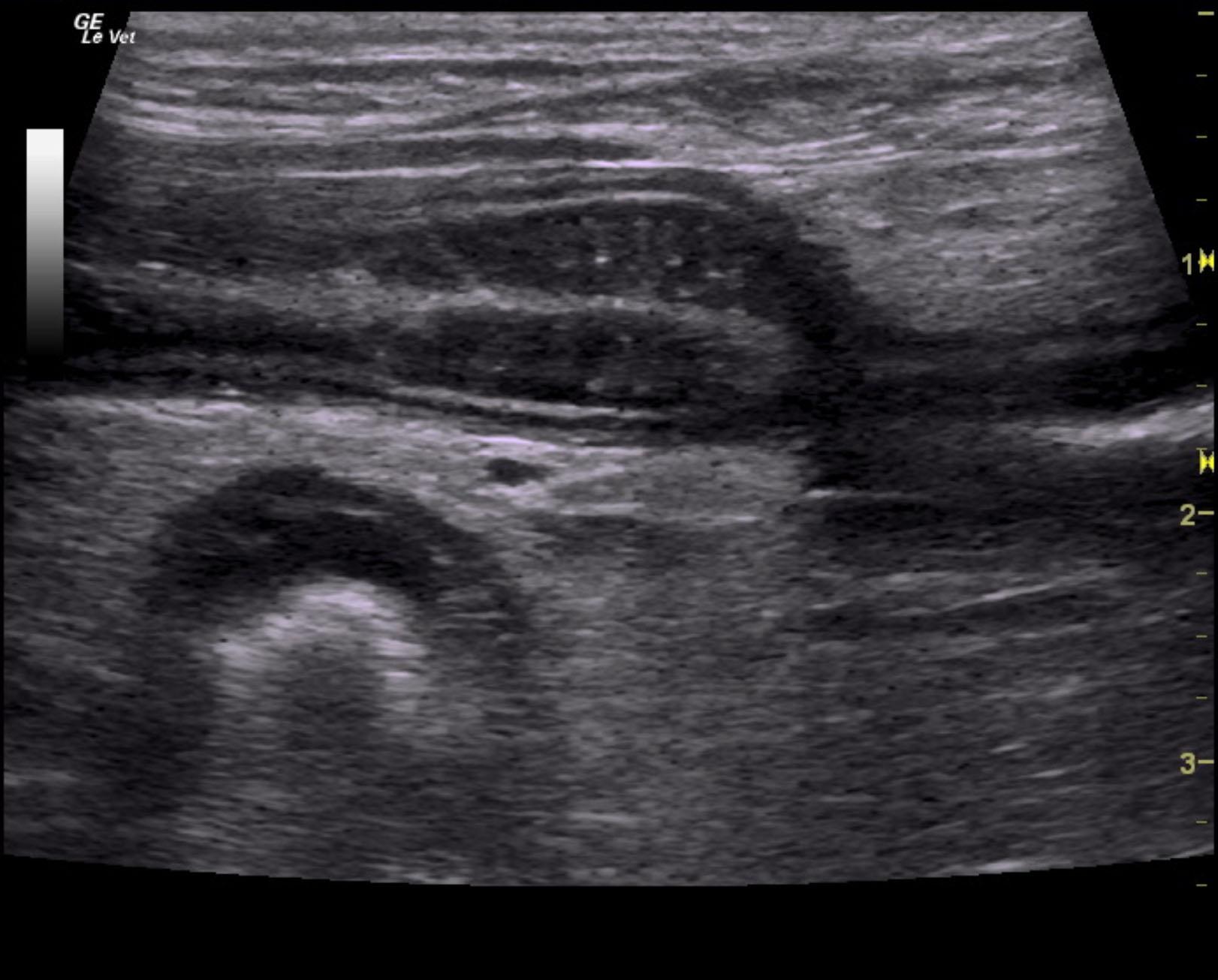
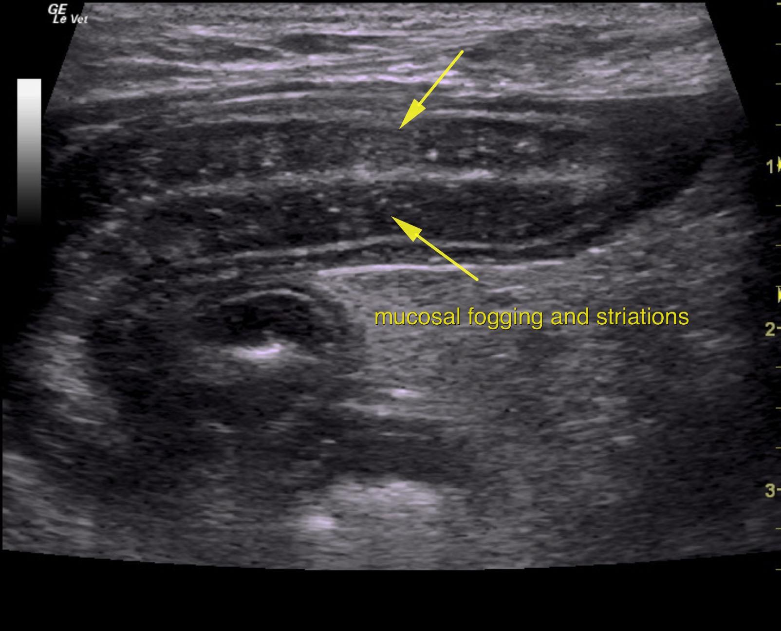
A 9-year-old SF Boston terrier was presented for evaluation of chronic diarrhea and weight loss. There had been no response to either metronidazole or Panacur. Urinalysis was normal. The only abnormalities on serum biochemistry were hypoproteinemia (3.6) and hypoalbuminemia (1.8).


Sonographic Differential Diagnosis

Reactive mesentery and pancreatitis pattern. IBD gastrointestinal pattern with mucosal fogging and striations. This is consistent with lymphangectasia.
Treatment for pancreatitis with plasma expanders and plasma transfusion would be ideal. Recheck sonogram is recommended in one week.

Image Interpretation

The pancreas revealed mixed, hypoechoic changes with hyperechoic surrounding fat. This is suggestive for concurrent pancreatitis primarily in the right limb in a region of 2 x 3 cm.
Slight free fluid was noted, likely owing to third spacing of fluid +/- lymphatic obstruction.
The gastrointestinal tract presented mucosal striations and fogging. This is consistent with lymphangectasia. Soft stool was noted in the colon.

DX

Pancreatitis

Outcome

None

Clinical Differential Diagnosis

GIT – IBD, lymphangectasia, dietary hypersensitivity, neoplasia, foreign body, intussusception

Sampling

None

Patient Information

Gender : Female, Spayed
Species : Feline
Type of Imaging : Ultrasound
Status : Complete

Clinical Signs

  • Diarrhea
  • Weight loss

Images

_20160227152615mag1mag3

Blood Chemistry

  • Albumin, Low
  • Total Protein, Low

Clinical Signs

  • Diarrhea
  • Weight loss
Skip to content