A 7-year-old spayed female cat was presented for vomiting and anorexia. On physical examination, thickened small intestine and a firm mass in the cranial abdomen was present. Abnormalities on CBC and serum chemistry included thrombocytopenia and hyperglobulinemia. Urine, fecal analysis, and T4 were all within normal limits. Cyproheptadine and famotidine was prescribed but the owner was unable to medicate.
A 7-year-old spayed female cat was presented for vomiting and anorexia. On physical examination, thickened small intestine and a firm mass in the cranial abdomen was present. Abnormalities on CBC and serum chemistry included thrombocytopenia and hyperglobulinemia. Urine, fecal analysis, and T4 were all within normal limits. Cyproheptadine and famotidine was prescribed but the owner was unable to medicate.
Case Study
Pancreatic suppurative inflammation in a 7 year old FS cat
Sonographic Differential Diagnosis
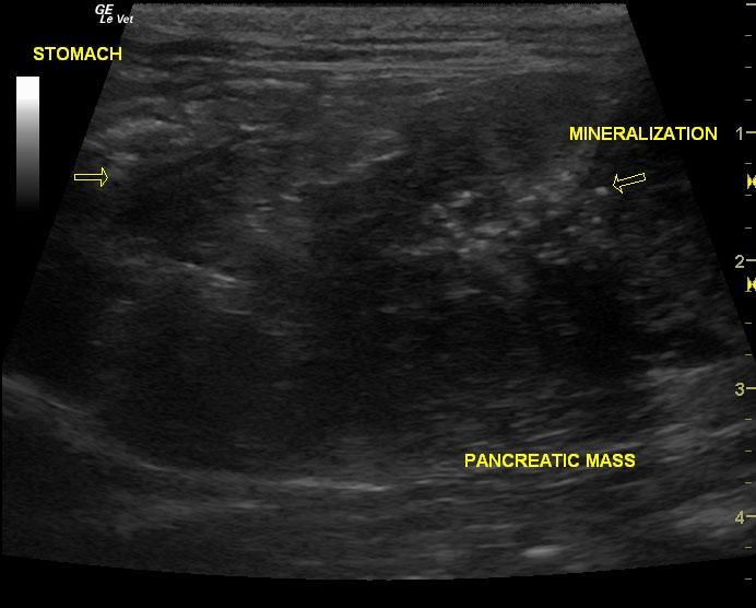
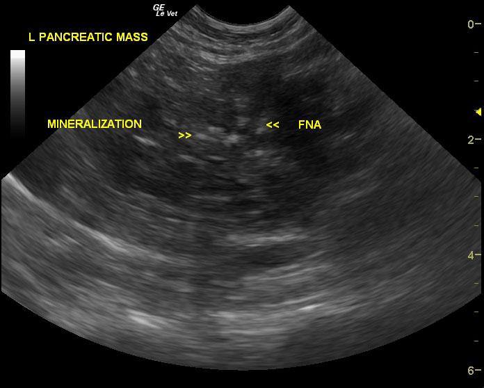
Mineralizing pancreatic mass. This presentation is most consistent with pancreatic carcinoma with a minor possibility of sequestrum and necrosis. The lesion does not appear resectable given the multiple lymph nodes involved. However, debulking effort can be considered depending upon the final cytology results. Remainder of the abdomen was normal.
Image Interpretation
The left limbs of the pancreas revealed a mixed, hypoechoic mass with multiple, focal hyperechoic mineralizing lesions and regional prominent hypoechoic lymph nodes. No free fluid was noted.
DX
Outcome
Metoclopramide was prescribed. It was noted that the cat was no longer vomiting a few weeks after the ultrasound.
Clinical Differential Diagnosis
Vomiting – GI tract pathology (obstruction, neoplasia, granuloma, IBD), pancreatic pathology (pancreatitis, neoplasia). Abdominal mass – neoplasia, granuloma, abscess or cyst of any of the following organs: liver, spleen, kidney, hydronephrosis. FIP.
Sampling
Ultrasound-guided fine needle aspirates of the left pancreatic mass was submitted to a laboratory. No complication from sampling was noted. The cytological interpretation of the sample revealed possible mild supportive inflammation.
Video
Patient Information
Clinical Signs
- Anorexia
- Vomiting
Exam Finding
- Palpable mass
- Thickened Intestines
Images
Blood Chemistry
- Globulin, High
CBC
- Platelet Count, Low
Clinical Signs
- Anorexia
- Vomiting