04_00524 Jazmin B

Case Study

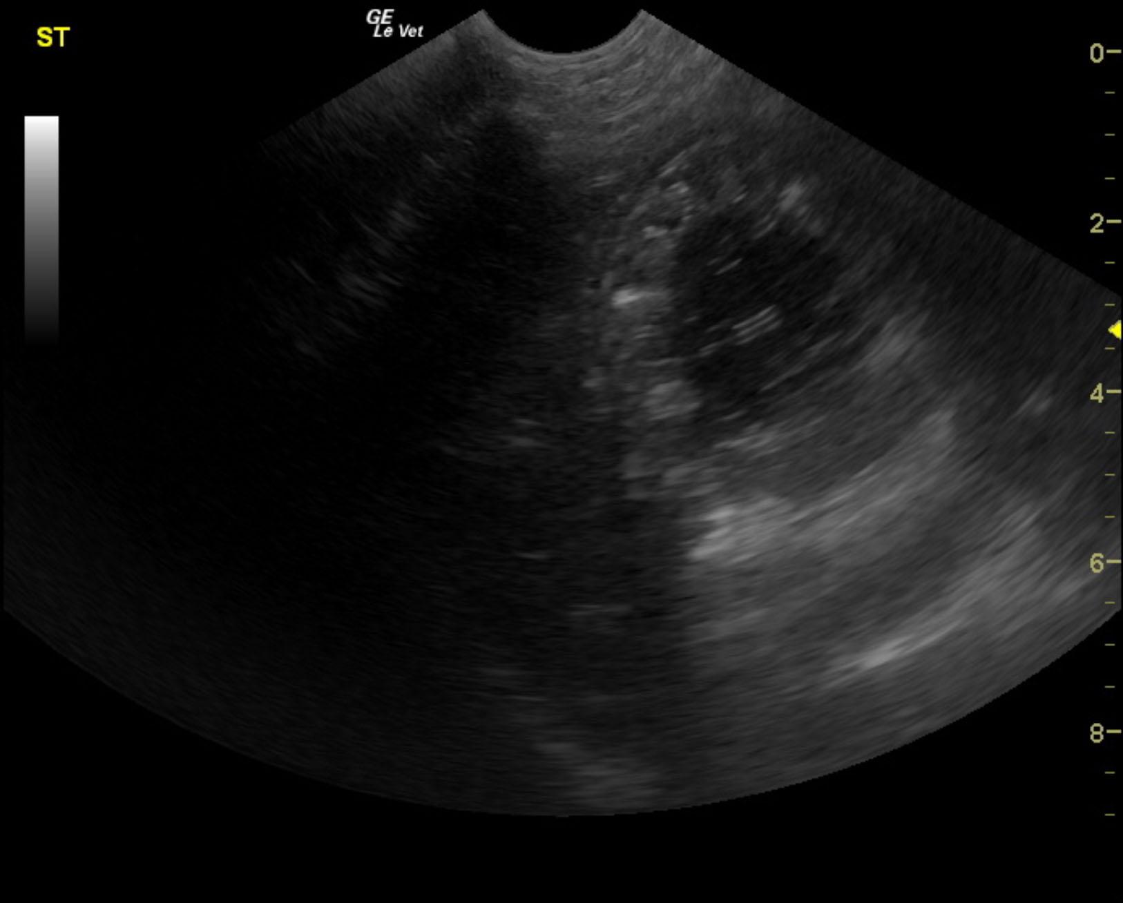
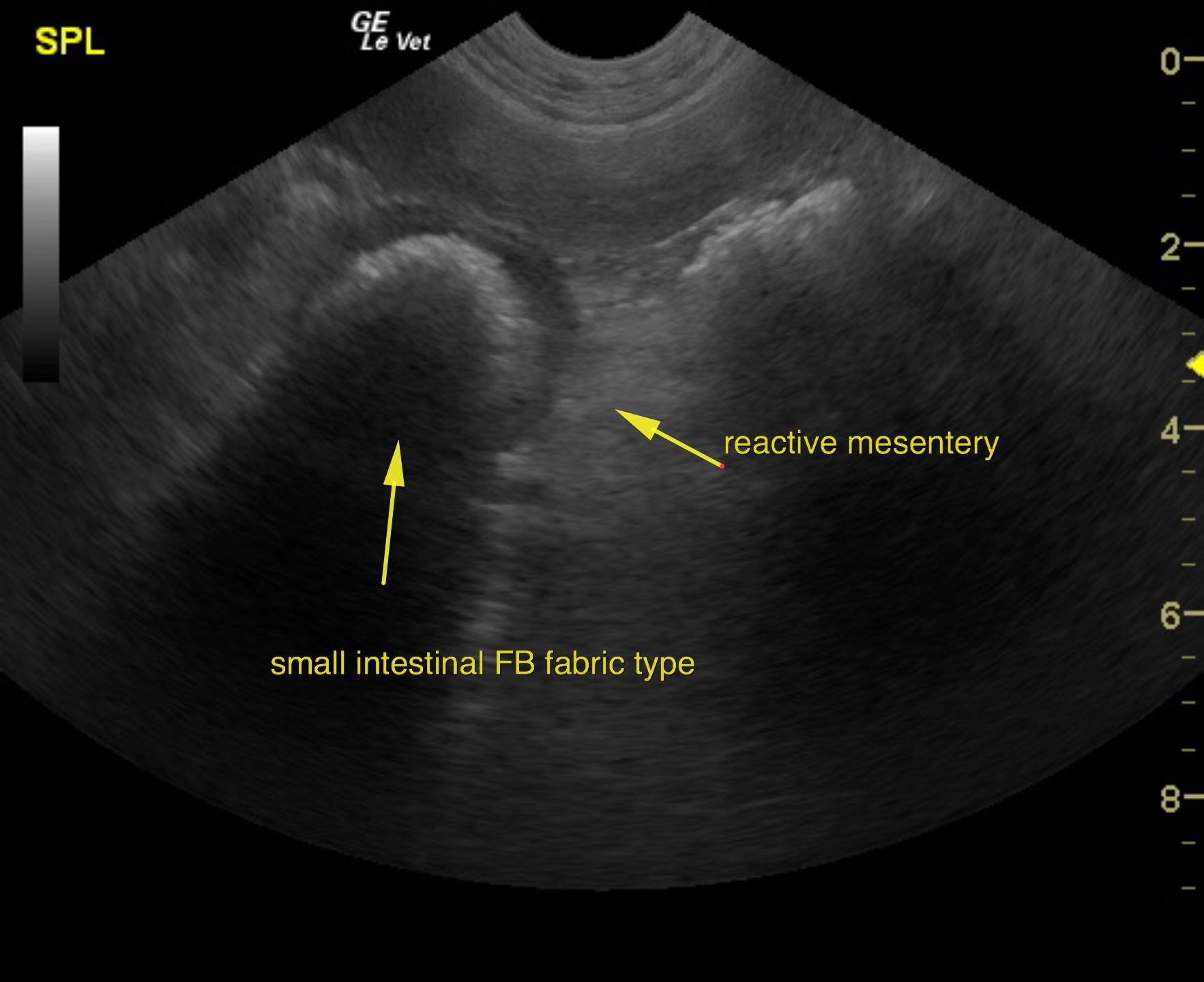
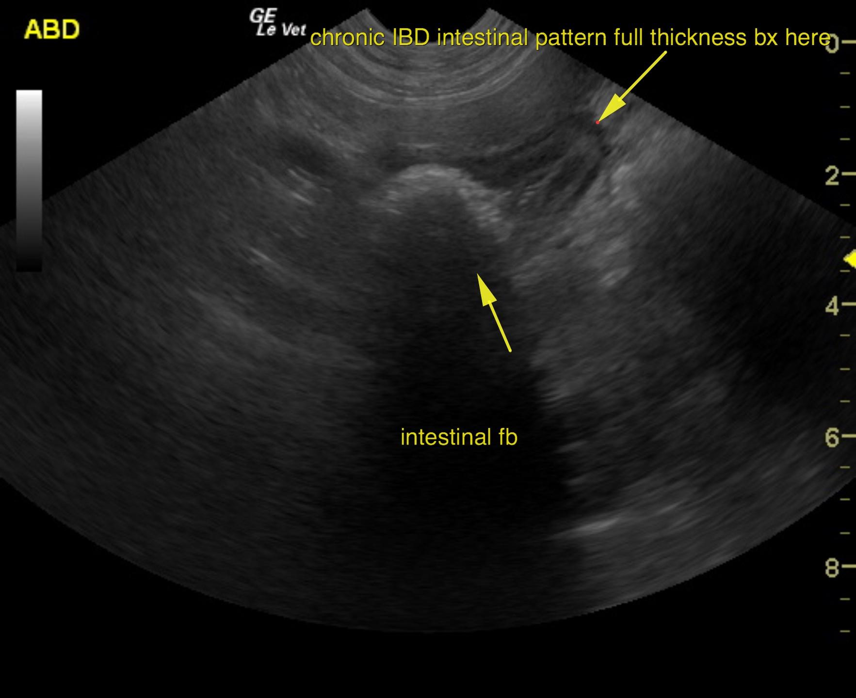
A 4-year-old SF Golden retriever with a history of previous previous foreign body surgery 3 months prior was presented for evaluation of vomiting. On survey radiograph colonic and small intestinal gas accumulation was evident. On CBC neutrophilia was present.  


Sonographic Differential Diagnosis

Distal small intestinal foreign body with thickened bowel and reactive mesentery.
Early emerging peritonitis.
Underlying inflammatory bowel disease or other intestinal disease is suspected.
Surgical intervention with expectations towards enterotomy and full thickness intestinal biopsies for long term management is strongly recommended in this patient. There is a minor potential for intestinal lymphoma.

Image Interpretation

The stomach was filled with fluid. Variable small intestinal mural thickening was noted in this patient. The distal small intestine revealed a shadowing foreign body that was followed by empty small intestine. This is consistent with fabric accumulation. Mural thickening was noted with reactive mesentery. Variable small intestinal mural thickening was noted with loss of detail. Intestinal biopsies are essential in this patient in addition to foreign body removal. A minor amount of free fluid was noted in the caudal abdomen. This is consistent with emerging peritonitis.

DX

IBD and foreign bodies

Outcome

Exploratory was performed. Resection and anastomosis of mid jejunum with removal of foreign body (pacifier) and 2 grass foreign bodies were removed. Biopsies were obtained at this time.

Clinical Differential Diagnosis

Non-specific gastro-enteritis (viral, bacterial, helminths, toxins, dietary indiscretion), foreign body, ulceration, IBD, neoplasia

Sampling

Intraoperative biopsies: 1. Jejunum (resected bowel segment): severe transmural ulcerative necrotizing enteritis, necrotizing steatitis of mesenteric fat with thrombosis.
2. Proximal jejunum (wedge biopsy): mild to moderate plasmacytic inflammatory bowel disease.
3. Duodenum (wedge biopsy): mild to moderate plasmacytic inflammatory bowel disease, hyperplasia of mucosal associated lymphoid cells (Peyer’s patches).

Patient Information

Gender : Female, Spayed
Species : Canine
Type of Imaging : Ultrasound
Status : Complete

Clinical Signs

  • Vomiting

Images

jaz1jaz2jaz3

CBC

  • Neutrophils, High

Clinical Signs

  • Vomiting
Skip to content