04_00516 Tuffy D Colonic stricture, cranial abd mass

Case Study

04_00516 Tuffy D Colonic stricture, cranial abd mass

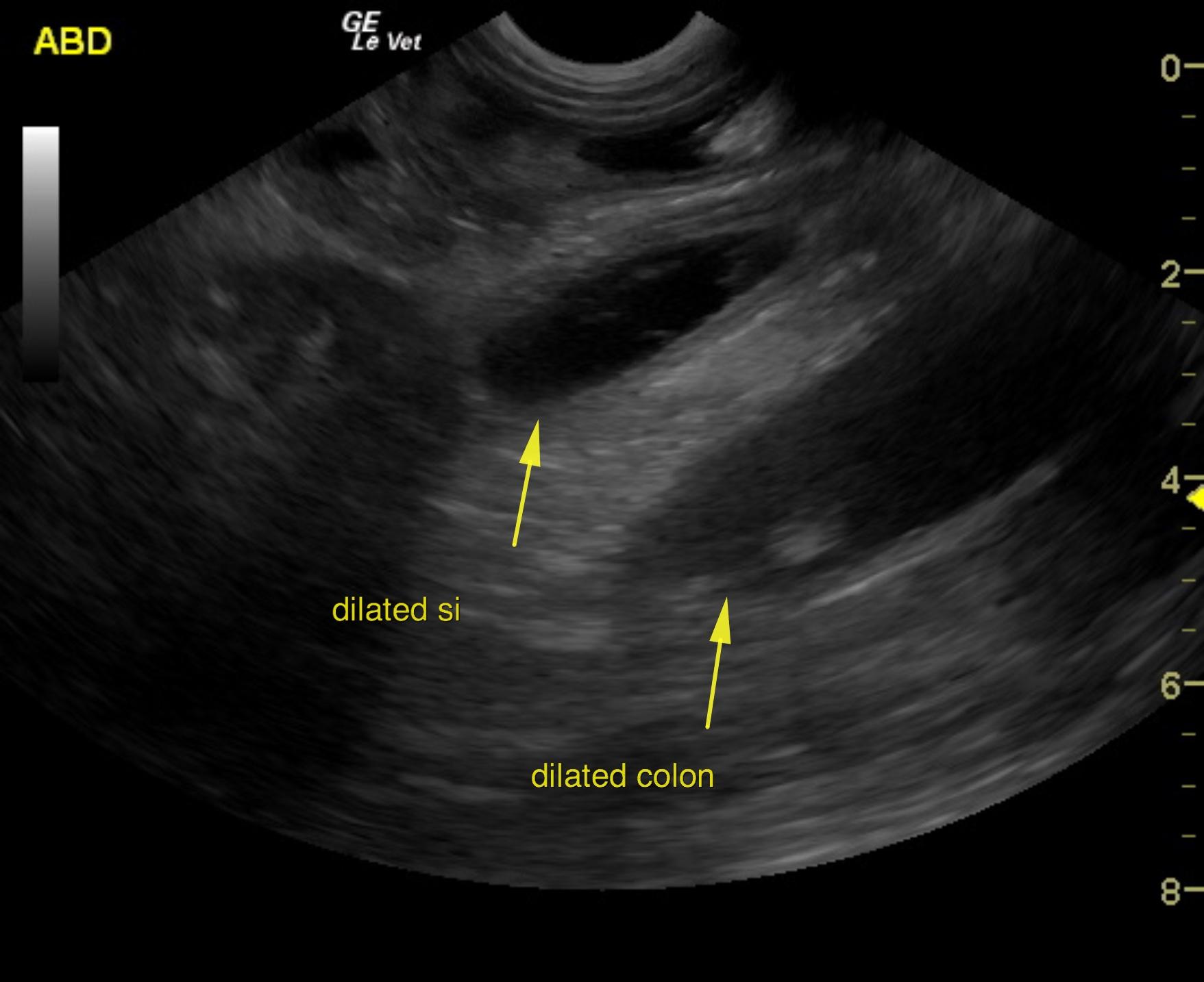
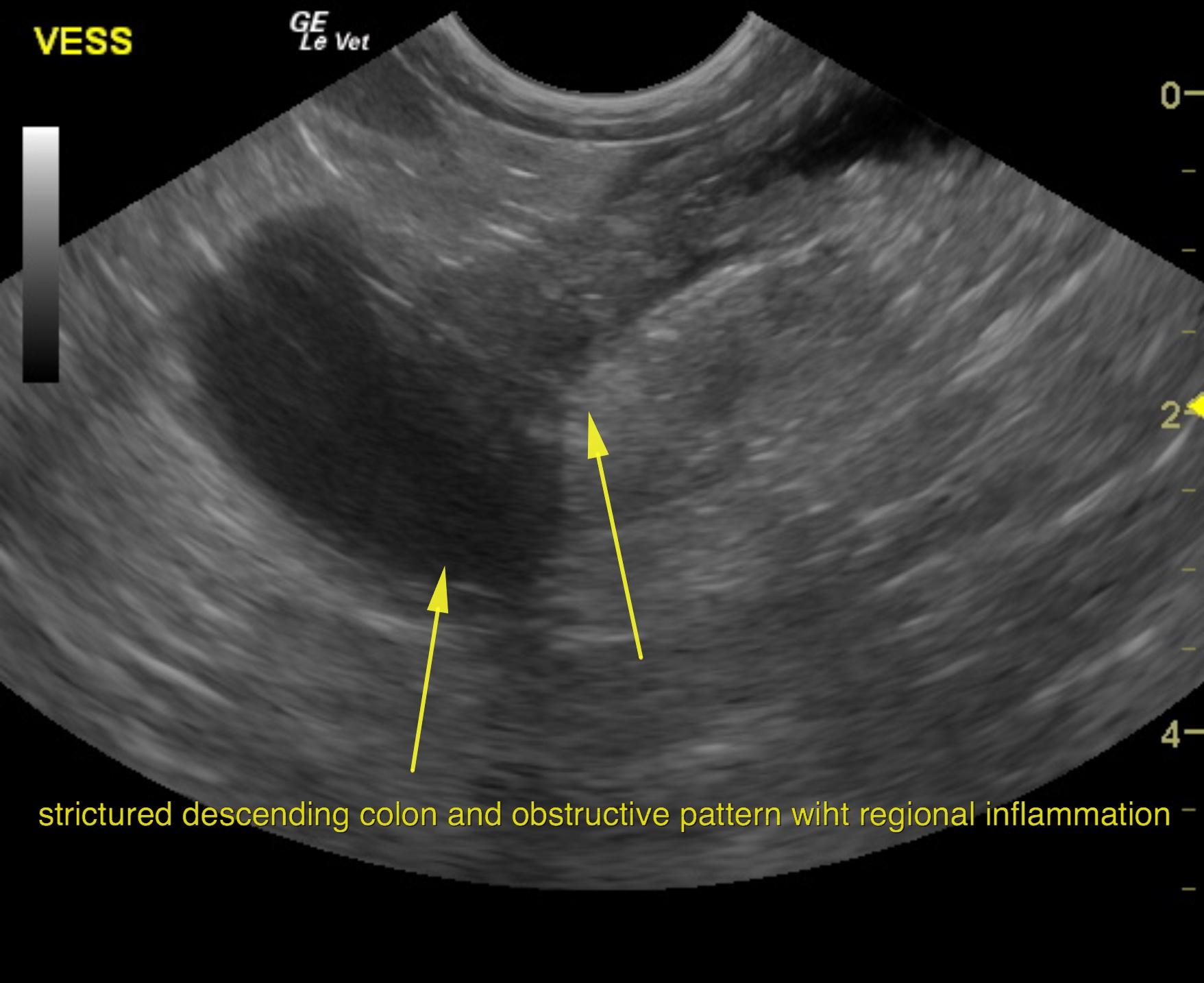
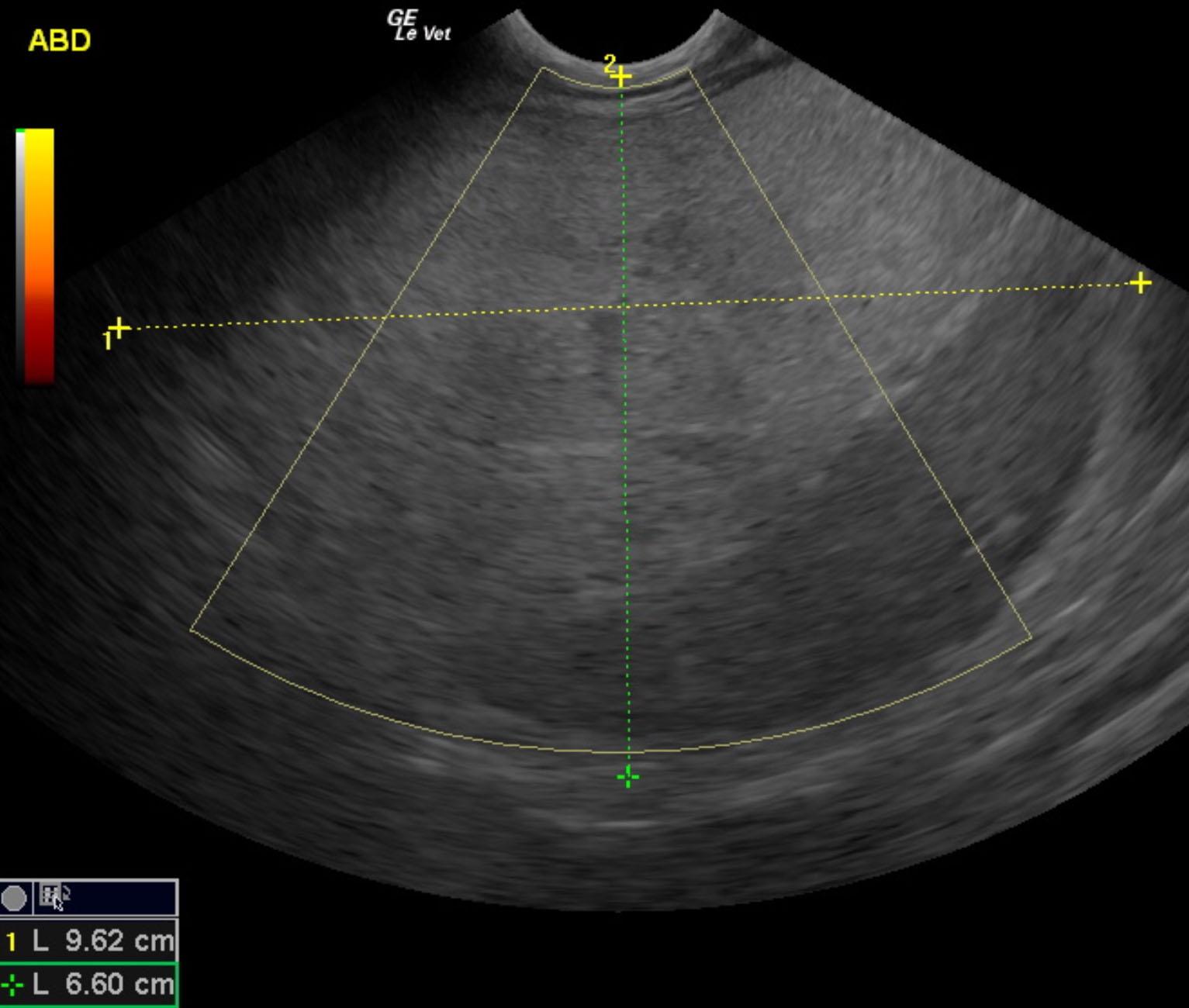
An 11-year-old NM Silky terrier was presented for evaluation of diarrhea and dry heaving/ crying during bowel movements. On survey radiographs splenomegaly and a cranial abdominal mass was evident. The only abnormality on serum biochemistry was hyperglobulinemia (5.3).


Sonographic Differential Diagnosis

Stricturing descending colon with diffuse intestinal bowel loop thickening or partial obstructive pattern. Cranial abdominal mass liposarcoma or omental abscess is suspected. This impinges upon the spleen.

Image Interpretation

The small intestine revealed dilated loops of bowel. Dilated intestine was noted with empty small intestine. Regional inflammation was present. The descending colon was thickened in this patient with a stricturing thickening with fluid accumulation prior creating an obstructive pattern. Regional inflammation was noted with diffusely thickened colon. The pattern is more consistent with colitis; however, underlying neoplasia is entirely possible given the loss of mural detail and stricturing pattern.
The abdominal mass appeared to be deriving from the pancreas or omentum. This was consistent with an omental abscess or liposarcoma or similar. Regional inflammation was noted. The remainder of the pancreas was unremarkable.

DX

Stricturing colonic thickening, cranial abdominal mass

Outcome

None

Clinical Differential Diagnosis

Splenomegaly – reactive, neoplasia, splenitis, torsion, congestion
Mass – neoplasia, abscess, cyst, granuloma of liver, kidney, pancreas, mesentery, lymph node

Sampling

None

Patient Information

Gender : Male, Neutered
Species : Canine
Type of Imaging : Ultrasound
Status : Complete

Clinical Signs

  • Diarrhea
  • Pain

Images

col2colon1tuf1

Blood Chemistry

  • Globulin, High

Clinical Signs

  • Diarrhea
  • Pain
Skip to content