A 13-year-old SF DLH was presented for evaluation of intermittent vomiting and abdominal discomfort. The only abnormality on serum biochemistry was mild hyperglycemia. Survey radiographs showed slightly thickened bowel loops.
A 13-year-old SF DLH was presented for evaluation of intermittent vomiting and abdominal discomfort. The only abnormality on serum biochemistry was mild hyperglycemia. Survey radiographs showed slightly thickened bowel loops.
Focal infiltrative intestinal pattern with obstruction of hairball or similar material. Regional inflammation. This is consistent with spontaneous necrosis, intestinal lymphoma, mast cell disease or dry form FIP.
IBD gastrointestinal changes noted elsewhere.
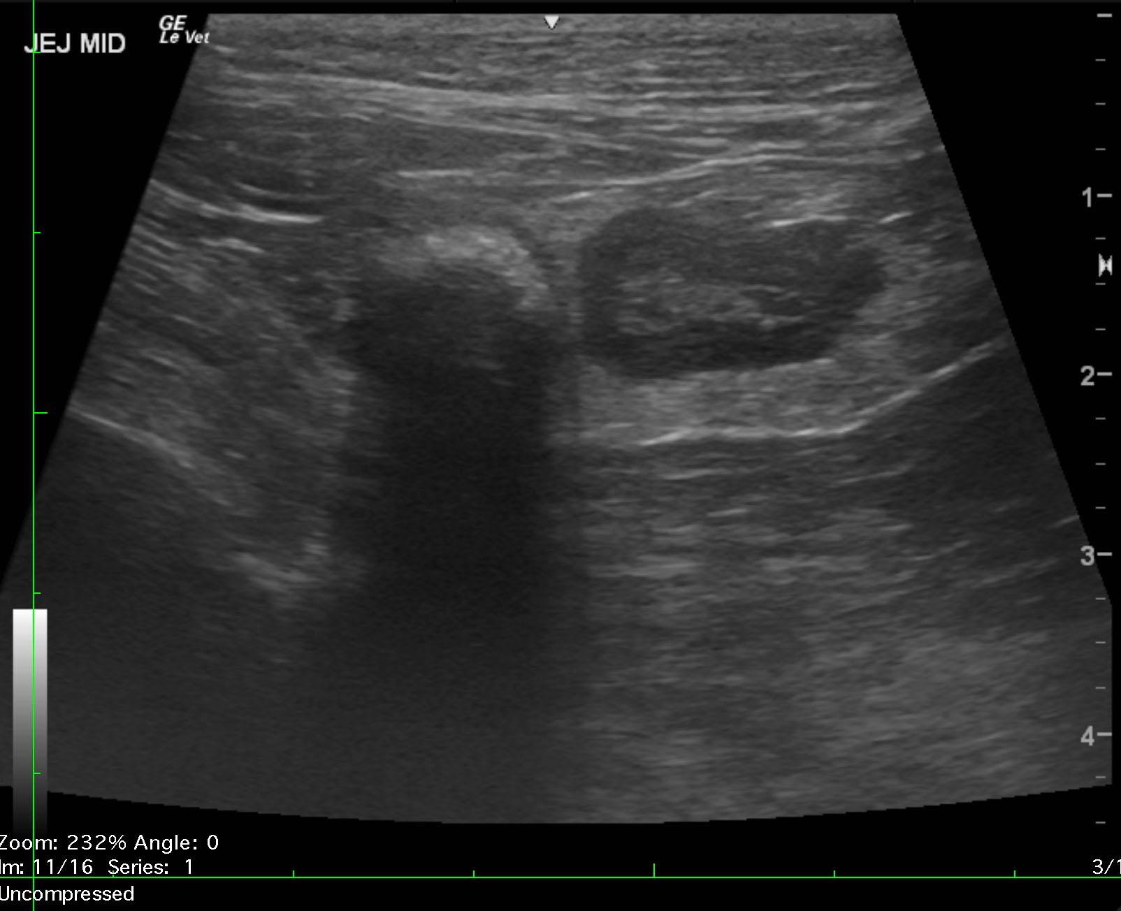
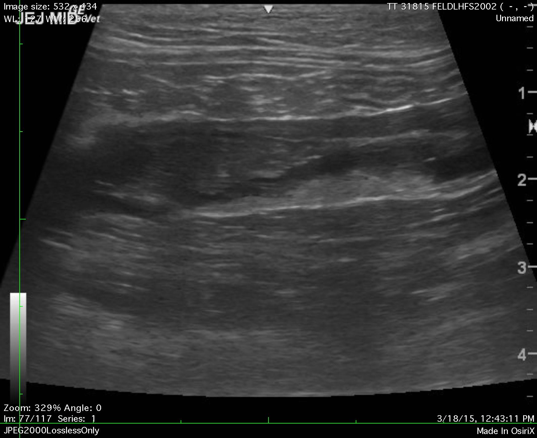
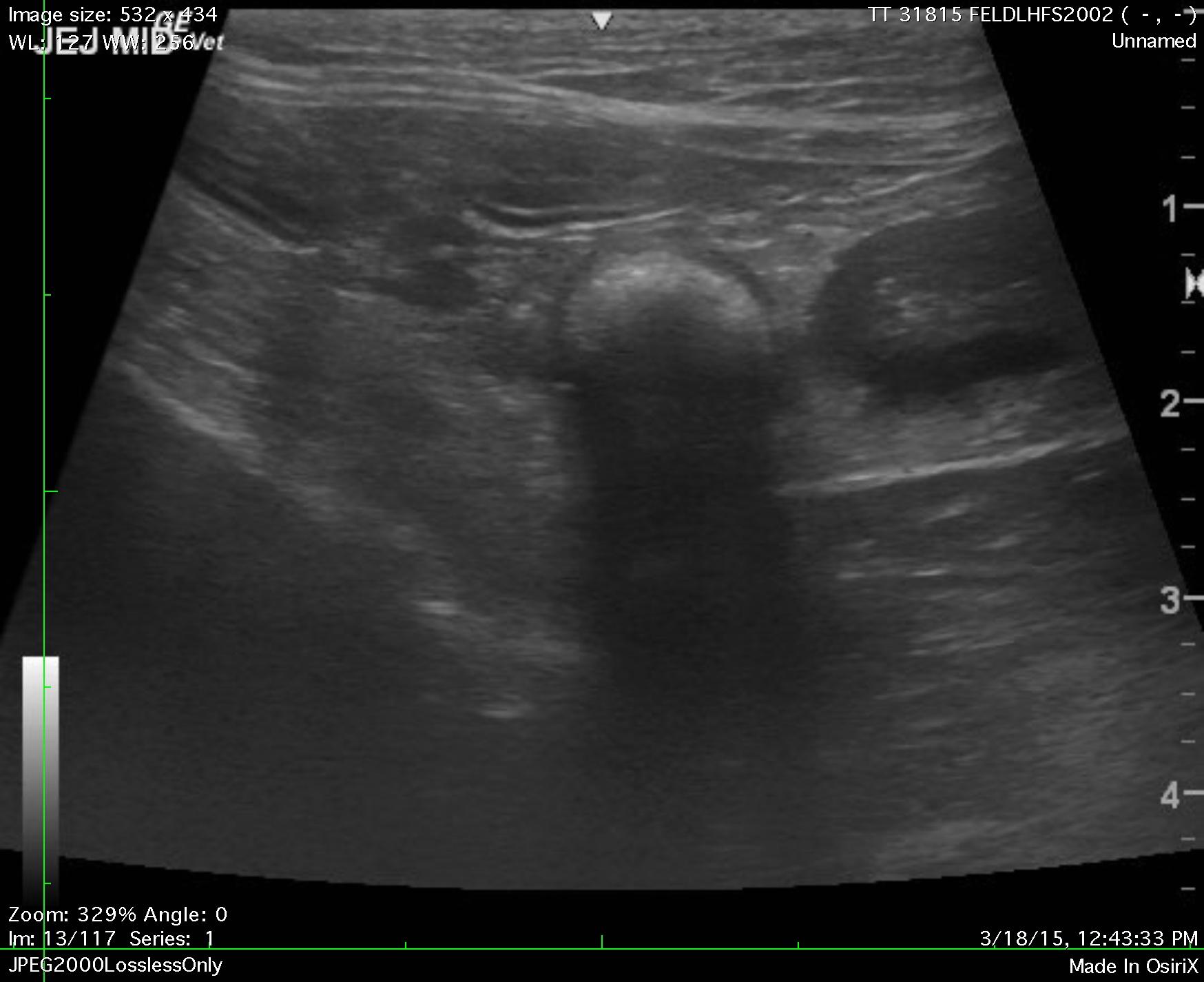
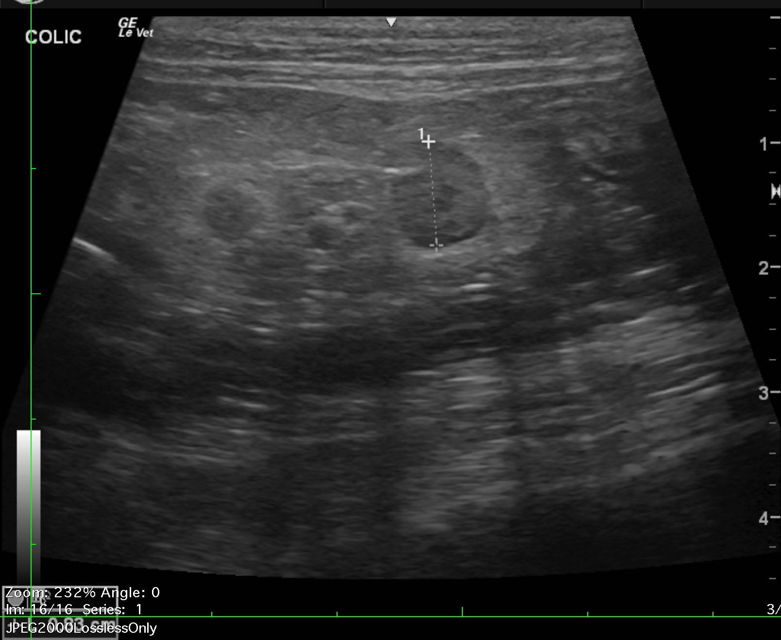
The gastrointestinal tract revealed mild, diffuse, hypertrophy of the muscularis with a 1:1 muscularis to mucosal ratio. The mid jejunum revealed thickened bowel that measured 0.5 cm serosa to mucosa with focal loss of detail. This extended for approximately 5.0 cm with echogenic chyme retention as well as regional, hyperechoic, surrounding fat attached to the serosa. This is suggestive for spontaneous perforation. Prior to the portion of infiltrative jejunum a hairball type density was noted. This may be a foreign body or passage of a hairball and is obstructed at the portion of infiltrative jejunum. The foreign object or hairball measured approximately 0.5-0.7 cm. The colic lymph node was enlarged and nodular with hyperechoic surrounding fat. This is suggestive for inflammation and measured 0.83 cm.
None
GIT – IBD, neoplasia, granulomatous enteritis, foreign body
Pancreas – pancreatitis, neoplasia
None