A 14-year-old NM DSH was presented for evaluation of chronic weight loss and grade II/VI systolic murmur.
A 14-year-old NM DSH was presented for evaluation of chronic weight loss and grade II/VI systolic murmur.
Gastric lymphoma or similar. A left medial cystadenoma type mass, possible adenocarcinoma. This is not likely responsible for the clinical signs.
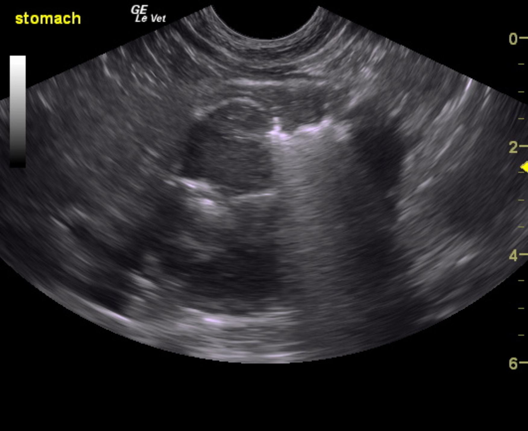
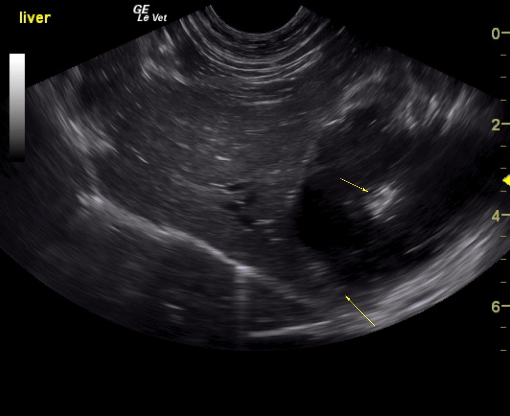
The gastric fundus was occupied by an infiltrative, hypoechoic mass that extended into the area of the esophageal inlet. The mass measured approximately 4.0 cm. The remainder of the intestinal tract was unremarkable including the pylorus which was free of evident pathology. The liver in this patient presented a microcystic and expansive, echogenic mass that measured 5.63 x 3.77 cm. This is consistent with biliary cystadenoma, possible adenocarcinoma. Core biopsy is necessary for a definitive diagnosis or surgical removal. The mass occupies the left medial liver and appears to be resectable.
None
Hyperthyroidism, neoplasia, IBD, cardiomyopathy, chronic kidney disease, chronic hepatopathy
None