04_00502 Tiki F Colonic mass with stricture

Case Study

04_00502 Tiki F Colonic mass with stricture

A 15-year-old SF DSH with a history of diabetes mellitus and recent onset suspected spinal vascular accident was presented for evaluation of acute diarrhea, decreased appetite, and abdominal distension. The only abnormality on urinalysis was glycosuria.


Sonographic Differential Diagnosis

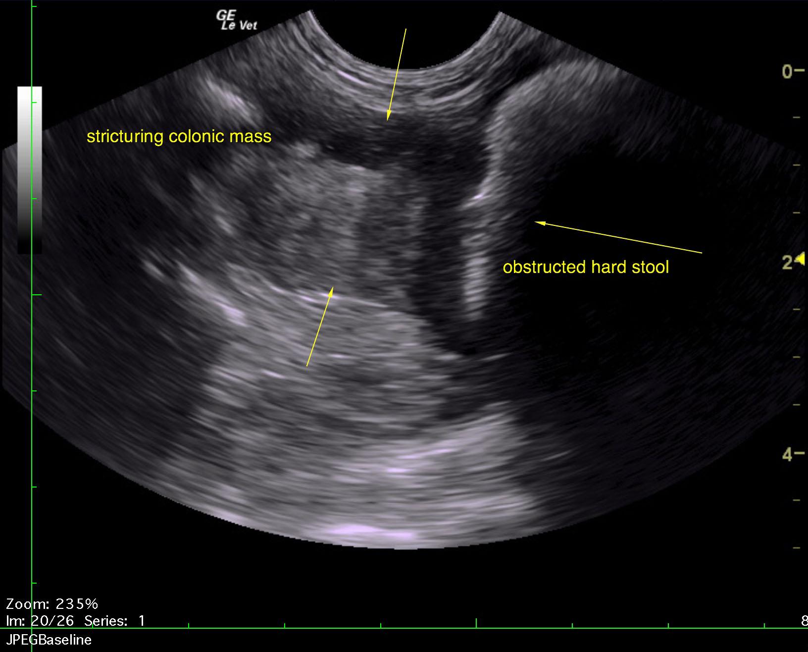
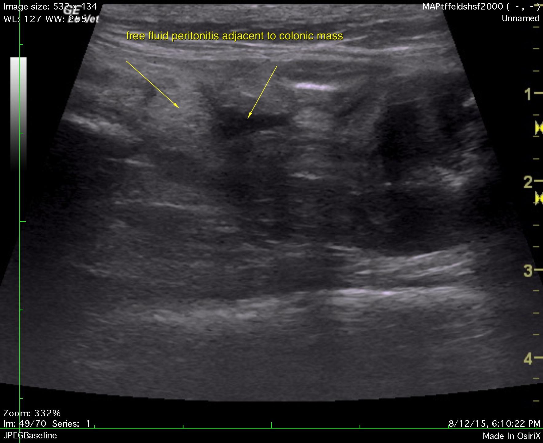
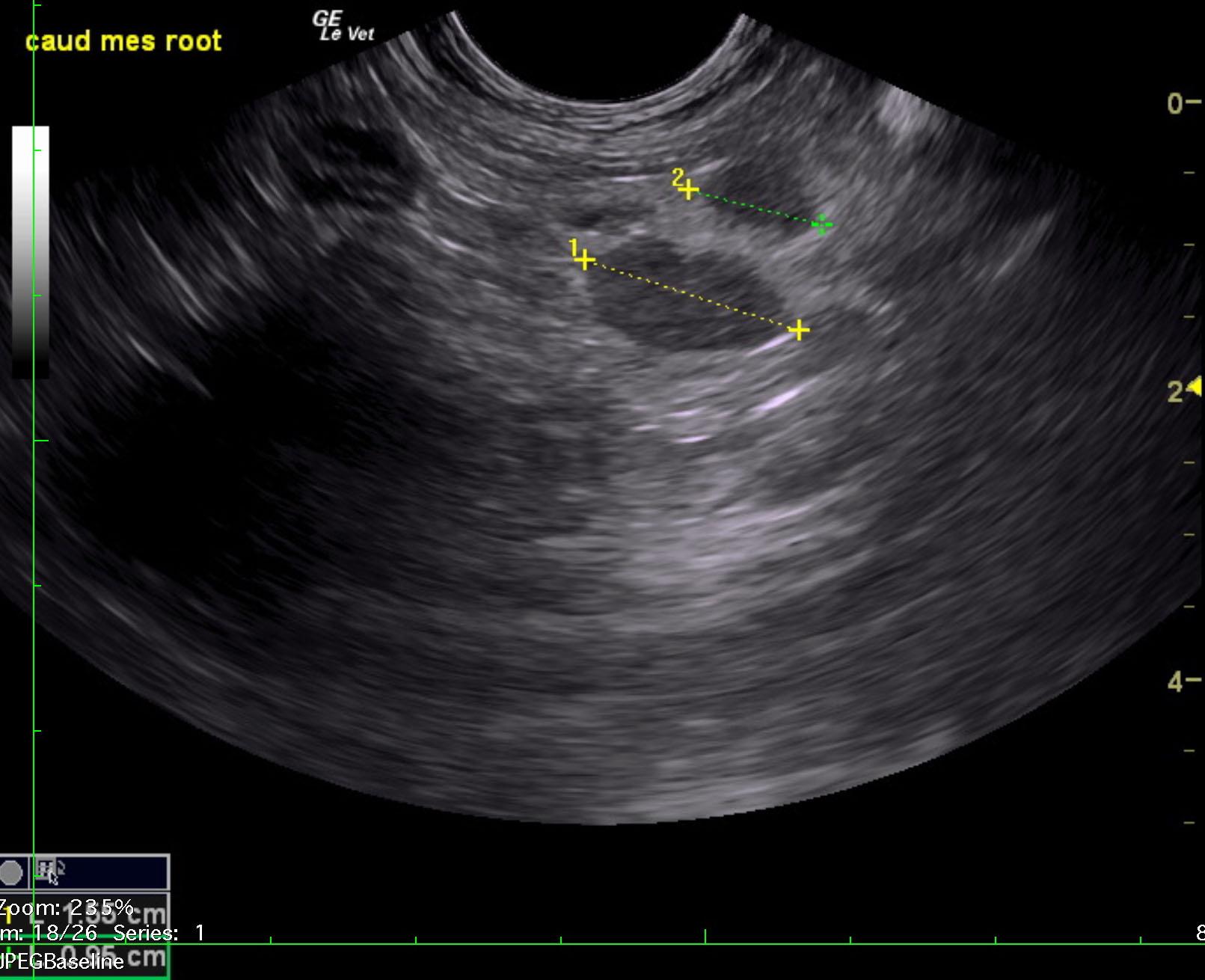
Stricturing colonic mass, obstructive pattern. Colic mesenteric lymphadenopathy with reactive fat. Trace amount of free fluid was noted adjacent to the colonic area. This is strongly consistent with a colonic carcinoma, possible lymphoma. Given the neurological issues metastatic disease should be a primary concern. CT with contrast of the CNS is recommended. Guarded to poor prognosis. Ultrasound-guided FNA of the mass could be considered for further definition to assess for potential chemoresponsiveness. Regional peritonitis was noted around the stricturing mass, possible perforation. Analysis of the free fluid is recommended.

Image Interpretation

The descending colon in this patient was significantly repleted. A stricturing colonic mass was noted and measured 2 x 4.0 cm. The mass was obstructive. Stool was impacted to the transverse colon. Peritonitis was noted around the stricturing mass. Regional lymphadenopathy was also noted and measured 1.5 x 1.0 cm each with reactive, hyperechoic ill defined fat. Mesenteric lymph nodes measured 1.55 cm and 0.95 cm with reactive surrounding fat.

DX

Colonic mass with stricture

Outcome

None

Clinical Differential Diagnosis

Uncontrolled diabetic
Metastatic neoplasia
Abdomen – septic peritonitis, FIP, neoplasia

Sampling

None

Patient Information

Gender : Female, Spayed
Species : Feline
Type of Imaging : Ultrasound
Status : Complete

Clinical Signs

  • Abdominal Distension
  • Anorexia
  • Diarrhea

Images

tiki_2tiki_3tiki_1

Clinical Signs

  • Abdominal Distension
  • Anorexia
  • Diarrhea

Urinalysi

  • Glucose Present
Skip to content