04_00454 Cassie T Strangulated hernia

Case Study

04_00454 Cassie T Strangulated hernia

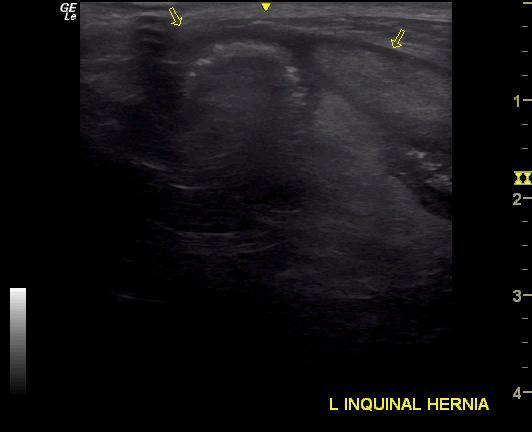
A 12-year-old SF Miniature Dachshund with a history of a left inguinal hernia, which had recently become harder and tender, was presented for evaluation of vomiting. Previous abdominal ultrasound had shown hepatic nodules, suspected to be benign.
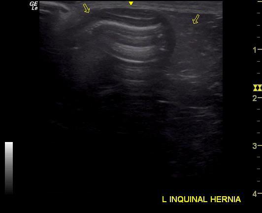
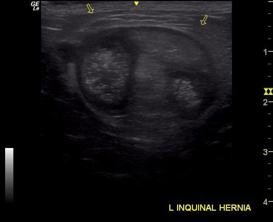
A 12-year-old SF Miniature Dachshund with a history of a left inguinal hernia, which had recently become harder and tender, was presented for evaluation of vomiting. Previous abdominal ultrasound had shown hepatic nodules, suspected to be benign.
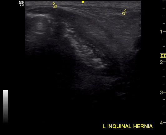
Sonographic Differential Diagnosis

Strangulated hernia with herniated small intestine and fat. Underlying mural intestinal disease. Possible underlying lymphoma. Variable other small intestinal wall thickening noted. Regional inflammation. Emerging peritonitis. Undefined nodular hepatic changes should be biopsied at surgery. Likely nodular hyperplasia, possible suppurative changes. Bilateral adrenal hypertrophy. Immediate exploratory surgery is recommended.

Image Interpretation

The small intestine appeared to be herniated along with fat into the body wall. Immediate surgical intervention is recommended. Regional inflammation associated with the herniated portion of the small intestine. Some transmural thickening noted. Resection and anastomosis likely will be necessary in the intestinal wall. If an intraabdominal approach is taken, then liver biopsy is recommended at the same time. Even though strangulation of the intestine is an issue, underlying mural disease appears to be an issue as well. Regional inflammation noted associated with other portions of small intestine in the abdomen should be investigated at surgery. Intestinal biopsies are essential in this case. Upper gastrointestinal wall thickening was also noted. No loss of detail.

DX

Strangulated hernia

Outcome

None

Clinical Differential Diagnosis

Strangulated hernia
Liver – neoplasia, granulomas, abscesses
Pancreas – pancreatitis, neoplasia

Sampling

None

Patient Information

Gender : Female, Spayed
Type of Imaging : Ultrasound
Status : Complete

Clinical Signs

  • Palpable Mass
  • Vomiting

Exam Finding

  • Palpable mass

Images

_20150430180455_20150430180434_20150430180416_20150430180347

Clinical Signs

  • Palpable Mass
  • Vomiting