A 9-year-old SF Collie was presented for evaluation of chronic vomiting over the last year with weight loss.
A 9-year-old SF Collie was presented for evaluation of chronic vomiting over the last year with weight loss.
A 9-year-old SF Collie was presented for evaluation of chronic vomiting over the last year with weight loss.
A 9-year-old SF Collie was presented for evaluation of chronic vomiting over the last year with weight loss.
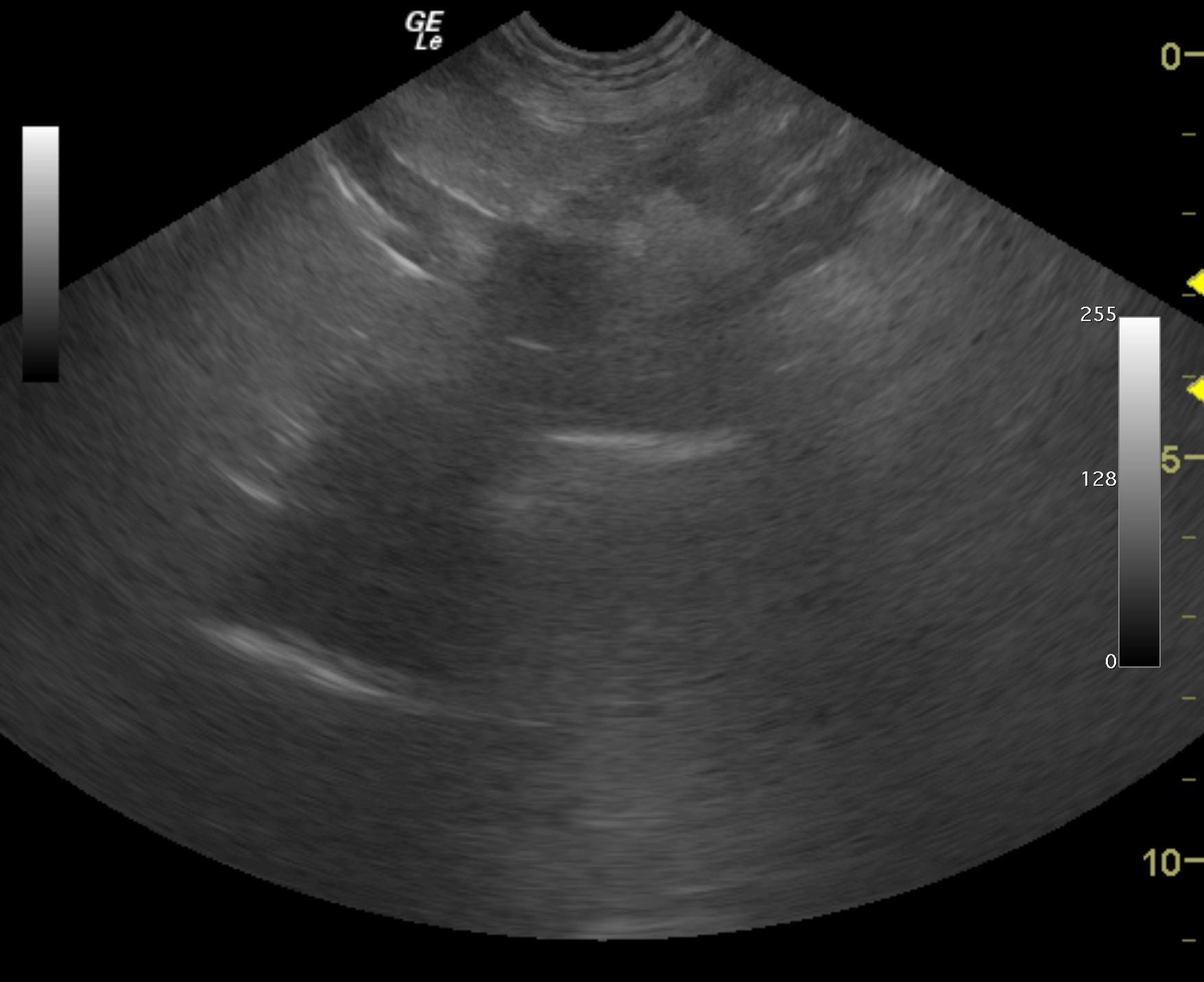
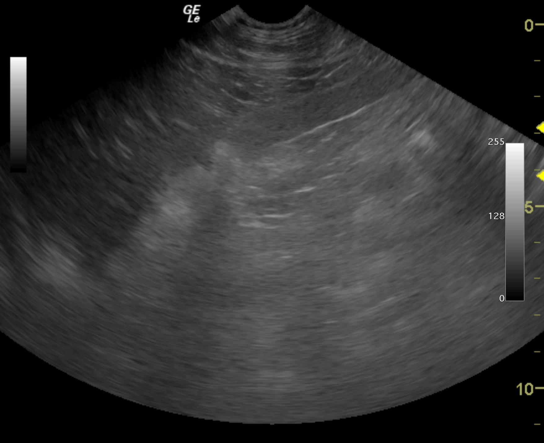
Gastric mass. Sarcoma such as lymphosarcoma, other round cell neoplasia, leiomyosarcoma or carcinoma are all possible. Granulomatous disease and non visible, embedded foreign body is possible. Ultrasound-guided FNAs were performed of the hypoechoic portion of the mass and submitted for cytology review. If no neoplasia is noted on aspirate results, then surgical exploratory is recommended with expectations towards debridement, assessment for any foreign body and appropriate surgical biopsies. I do not feel that the lesion can be cleanly resected given the concentric manner in which it is formed and enters into the gastroesophageal inlet.
The gastric fundus was significantly thickened. This created a hypoechoic mass type structure for 5-6 cm. However, this may represent granulomatous disease even if neoplastic criteria is met. The pylorus was largely unremarkable and the majority of the pathology was associated with the body of the stomach and gastroesophageal inlet with surrounding omentum and pancreas. Gastric wall thickness from lumen to serosa was approximately 2-2.5 cm. The gastric fundus was hypoechoic with mixed echogenic hyperechoic changes around it. Complete loss of detail and a mass type structure that measured 5 x 6 cm entering into the gastroesophageal inlet was noted. Clean view of the gastroesophageal inlet was not possible. FNAs of the hypoechoic portion of the gastric wall were performed without complication.
None
GIT – neoplasia, IBD, ulceration, foreign body, intussusception, granulomatous enteritis, dietary hypersensitivity
Chronic kidney disease
FNA of the gastric mass revealed carcinoma.